Showing posts with label Open Access Journal of Complementary & Alternative Medicine. Show all posts

Friday, August 19, 2022

A Reflection on Yoga, Acupuncture and Medicine


Introduction

In the 20th Century a great effort has been made both to prove that yoga and acupuncture (i.e., Chinese medicine) can be used for health-related purposes and to discover the functional mechanisms of both systems. From the Western point of view, when both yoga and Chinese medicine became popular as a Complementary and Alternative Medicine (CAM) a resistance became perceptible that led to regulation of CAM (CAMDOC Alliance, 2010). The main issue is that Western and Eastern medicine have a different approach to both health and treatment of illness.

In case of yoga, it has to be stated that it was not conceived as a medical system, but as a system for the discovery of the ultimate goal of humans – Self-realization. Thus, its methods – though there are a great number of yoga traditions – are directed to this aim. Nevertheless, hatha yoga has a strong emphasis on health and has a wonderful spectrum of procedures for arriving at a health status that enables intensive pursuing of the said ultimate goal manifested as samadhi that follows from the cessation of the activities of chitta – the substance of mind/mindfield. The result stated in Patanjali Yoga sutra (PYS) is:


Read more about this article :https://lupinepublishers.com/complementary-alternative-medicine-journal/fulltext/a-reflection-on-yoga-acupuncture-and-medicine.ID.000155.php

Read more Lupine Publishers Google Scholar Articles : https://scholar.google.com/citations?view_op=view_citation&hl=en&user=0sWAIssAAAAJ&citation_for_view=0sWAIssAAAAJ:a0OBvERweLwC

Friday, July 29, 2022

Detection and Analysis of Pathogenic Bacteria from Raw and Pasteurized Milk


Abstract

Milk is a proper and complete healthy diet for all individuals. Milk contains all the basic nutrients such as carbon, calcium, and rich water content which facilities bacterial contamination if hygienic conditions are not properly maintained. Different types of bacteria such as gram-positive and gram-negative can contaminate milk in many ways such as use of unclean utensils, storage tanks, and other factors which includes diseased animals, or/and animals suffering from mastitis. Most commonly isolated organisms from raw milk samples are E. coli, Shigella, Staphylococcus aureus, Klebsiella, and spores forming Bacillus species which can cause serious enteric illness if contaminated consumed. Pasteurization process extensively reduces the risk of contamination and makes the milk safer for drinking. In this study, we have collected 24 samples in total of raw milk (n=12) and pasteurized milk (n=12). Further, we processed the milk samples on different agars. For raw milk, we used Nutrient agar, Mannitol salt agar, and Eosin Methylene blue agar whereas for pasteurized milk we have used dye reduction test. Raw milk indicated high contamination as compared to the milk which is pasteurized. Even some locally available pasteurized milk has detected contaminating agents. As the results indicated that raw milk is highly contaminated so for the safety of individuals and the population, it is suggested that we should use pasteurized milk in our daily life. Furthermore, we must apply some safety precautions including maintenance of tanks, storage temperature, and sterilization of utensils in which milk is kept.


Read more about this article : https://lupinepublishers.com/complementary-alternative-medicine-journal/fulltext/detection-and-analysis-of-pathogenic-bacteria-from-raw-and-pasteurized-milk.ID.000151.php

Read more Lupine Publishers Google Scholar Articles : https://scholar.google.com/citations?view_op=view_citation&hl=en&user=0sWAIssAAAAJ&citation_for_view=0sWAIssAAAAJ:4OULZ7Gr8RgC


Friday, September 18, 2020

Lupine Publishers | Diversity of Food-Borne Pathogens Isolated from Raw Meat Sample

 Lupine Publishers | Open access Journal of Complimentary and Alternative Medicine


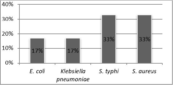
Foodborne diseases are considered to be the wide-spread health problem in most parts of the world especially in developing countries. The aim of the study evaluates the frequency of microbial contamination of raw meat that is often contaminated with foodborne pathogens. The perishable nature of nutritious meat is one of the potential sources of pathogens as well as an excellent vehicle for transmitting foodborne disease in human beings. In this study, Six (n=6) samples of meat such as beef, mutton, lamb, chicken, fish, camel meat were collected from common retail shop of Karachi and analyzed for the prevalence of pathogenic microorganism by using standard plate methods to purify it on specific agar and to perform biochemical testing for identification and isolation of bacterial species. In our study, it was found that 33% isolates of S. typhi and S. aureus were found whereas 17% isolates of E. coli and Klebsiella pneumoniae were detected from various meat samples. It is therefore expedient that great care should be taken during handling and good hygiene practices should be followed to reduce the microbial load to a harmless level.

Keywords: Food; Disease; Organisms; Meat sample

Introduction

Meat has long been known for its enrichment composition containing a rich source of protein, carbohydrate, vitamins, and fats. Most people rely on meat due to the presence of this entire rich nutrient that is required for the human body. Meat including beef, mutton, poultry, fish, lamb, and camel meat [1].

Nutritional composition of beef and mutton and their role in the human immune system

Meat is one of the most edible and delicious food in the world that play a role in human metabolic functions. Beef and mutton meat contain a large amount of protein and Vitamin (B12, B3 (Niacin), B6), Iron, Zinc, Selenium, and minerals. This could be beneficial to the body for the building of body tissues and help in the repair mechanism. It also helps in the production of antibodies to build up immune system [2].

Composition of fish with the supplement of Omega 3 and their role in the human body

Proteins are the second major part of a fish constituent. Two essential amino acids called lysine and methionine are generally found in high concentrations in fish. Fish meat is also a valuable source of minerals such as sodium, potassium, magnesium, phosphorous, iron, and omega 3 fatty acids that can help in reproduction and brain development. Fish also has non-protein nitrogen (NPN) fraction which is water-soluble. The constituents of the NPN fraction play a major role in the quality of fish [3]. Raw fish is often contaminated with foodborne pathogens. It provides a perfect medium for the growth of the microbial pathogens such as Salmonella, Escherichia coli, Shigella sonnie and Listeria monocytogenes. They have been found in raw fish through washing water or may be from contact surfaces [4].

Composition of lamb meat and their function in the human body

Lamb meat is an excellent source of Vitamins B6, B12, protein, zinc phosphorous, selenium, folate, and choline that act as an antioxidant that prevents reactive oxygen species in the body. They also reduced the level of unwanted accumulation of homocysteine in the body. High blood levels of homocysteine responsible for causing cardiovascular disease. Another property of grass feed lamb meat contains Conjugated linoleic acid (CLA) that helps in reducing inflammation and body extra fat. The combination of both can reduce the risk of heart diseases [5]. Lamb meat can be contaminated with a variety of microorganisms such as Staphylococci, Corynebacterium, Streptococci, Micrococcus, Salmonella, Escherichia coli, coliform and Yeast [6].

Composition of fatless camel meat used for medicinal purpose

The demand for camel meat is mainly due to the health benefit purposes. It is a source of high-quality meat due to the presence of unsaturated fatty acids like linoleic acid which prevents from tumor or cancerous diseases. It also contains less fat content which is recommended for weight loss and prevent from heart diseases and atherosclerosis. It also reduces cholesterol level in the blood [7,8].

Composition of chicken and spread of microorganism in poultry meat

Chicken meat is one of the most consumed food in Pakistan. It is nutritious with rich protein content. All vitamin B are present in chicken meat, including B1, B2, B3, B5, B6, B12, folate, biotin, choline, selenium, zinc, copper, and omega 3. Recent research has proved that intake of chicken meat can reduce the risk of colorectal cancer. It contains fiber that can lower blood cholesterol and blood LDL-cholesterol [9]. Poultry meat is contaminated with a variety of microorganisms, including those capable of spoiling at freezing temperatures. Raw chicken meat is contaminated with certain foodborne pathogens such as Campylobacter jejuni, Salmonella enteritis, Staph. aureus, Listeria monocytogenes. Others may also be present which includes the most recently reported Arcobacter and Helicobacter spp and occasionally, verotoxigenic Escherichia coli [10].

Spoilage of Meat

The perishable nature of meat is highly sensitive to spoilage by contact with different microorganisms through exogenous and endogenous sources. Out of which, the intestinal tract of animal is the main source of microorganism. Organism that may infect a living animal causing endogenous diseases may also spoil the meat. Consumption of this meat from an infected animal can cause diseases in human. Three sources of exogenous contamination are by slaughtering, handling, and processing. Due to these biochemical changes that allow microorganisms to grow in meat and carry the greater chances of contamination [11]. Microbial spoilage of food is an area of global concern, causing serious foodborne intoxications and resulting in high economic losses for the food-producing sector.

The predominant bacteria associated with spoilage of meat are Carnobacterium spp., Enterobacteriaceae, Lactobacillus spp., Leuconostoc spp., Pseudomonas spp. and Shewanella putrefacients [12]. The poikilothermic nature of fresh fish meat that allows a wide variety of bacteria to grow and spoiled the fish which includes Gram- negative, rod-shaped bacteria which belong to the genera Pseudomonas, Moraxella, Acinetobacter, Shewanella, Flavobacterium, Aeroemonadaceae, and Vibrionaceae, and Grampositive bacteria such as Bacillus, Micrococcus, Clostridium, Lactobacillus, and Corynebacterium [13,14].

Mechanism of rigor mortis occurring in meat

Rigor mortis is a physiological change that occurs after the death of an animal in which the hardening of skeletal muscles becomes maintained for a certain period approximately within 12 to 24hrs. then gradually flexible after 24 hours. Normally glycogen present in the muscles of the animal. When the animal is slaughtered in the excited state, its glycogen converts into lactic acid in bulk amount. Suddenly the change in pH is occurred which turns into acidic so it allows acidophilic bacteria to grow which can spoil the meat easily. The process is known as rigor mortis occurring in meat [15]. Greater chances of bacterial contamination during slaughtering and then cutting by using tools such as knives, wooden board, weighing scale and meat mincer. Or it may the meat handler who is shedding microbes and can act as a carrier.

Presence of foodborne pathogens in meat through contact surfaces and processing

The perishable nature of meat provides a substrate to support the growth of microorganisms in meat. Meat contains a wide range of pathogenic microorganism that is obtained either from contaminated surfaces or during processing. Most of these bacteria can produce biofilms that protect them from external harm and enable them to adhere strongly to contact surfaces. The greater chances of bacterial pathogens during meat-processing and their equipment so the surfaces and processing both may contribute to the contamination of meat [16].

Bacterial Foodborne Diseases

Foodborne diseases are a major cause of death in developing countries by representing an important public health problem worldwide. It is assumed that foodborne and waterborne diarrheal diseases kill more than 2.2 million people each year. Foodborne diseases resulting from the ingestion of bacteria, toxins, and cells produced by microorganisms present in food. Toxins may not alter the appearance, odor, or flavor of food but may cause food poisoning. Common kinds of bacteria that produce toxins include Staphylococcus aureus and Clostridium botulinum. The intensity of the signs and symptoms may vary with the amount of contaminated food ingested and susceptibility of the individuals to the toxin. Foodborne microorganisms can cause severe illnesses to humans which may also lead to the drug resistance [17,18].

Approximately 69% of gram-negative bacteria are known to cause bacterial foodborne diseases. Several researchers have reported that the meat samples contaminated with a higher load of Klebsiella pneumoniae, Enterobacter spp, Pseudomonas aeruginosa, E. coli, Salmonella sp, Serratia marcescens, Proteus Vulgaris, Staphylococcus aureus and Bacillus sp. On the other hand, foodborne pathogens can disseminate from contaminated meat to the surfaces and can spread infections in the community [19-23].

Antibiotic Resistivity

Some bacteria have developed resistance against antibiotics that are not killed or controlled by antibiotics even they can multiply in the presence of these drugs. It could be more difficult to treat people if they became ill with the antibiotic-resistant strains such as the most recently reported resistance is carbapenem-resistant Enterobacteriaceae (CRE). These strains have developed resistance against carbapenem drug e.g. Klebsiella. Staph. aureus is found to be resistant against multi drugs and falls in the group of Methicillinresistant Staphylococcus aureus (MRSA), Salmonella typhi is found resistant against chloramphenicol, ampicillin, tetracycline, and trimethoprim. They are responsible for a well-known public health problem [24].

Materials and Methods

Sample collection

A total of Six (n=6) samples of raw meat were collected in a sterile container which includes minced chicken, beef, mutton, lamb, camel, and fish. 1 gram of each minced meat samples was weighed and transferred into a tube containing 9ml of selenite broth for enrichment. Then it was incubated at 37 °C for 24 hours. After incubation, streaking was done on nutrient agar plate. And the plates were incubated at 37 °C for 24 hours to the study was carried out to determine the microbial load among different raw meat available in common retail shops of Karachi, Pakistan. This study is designed to characterize the foodborne pathogens, which include S. aureus, E. coli, Salmonella, and Klebsiella isolated from raw meat samples and to determine the susceptibility pattern of isolated bacterial strains against the frequently prescribed antibiotics. These isolated bacterial strains are known as foodborne pathogens resulting in food intoxication. This research is necessary to the public which will create awareness among the consumers to prevent foodborne illnesses. Observe the colonial characteristics on nutrient agar and to perform gram staining and further identification tests to identify the isolates.

Antibiotic Susceptibility Testing

Antibiotic susceptibility testing is performed by using Kirby- Bauer disc diffusion method in which loop full culture of the identified isolate was streaked on MHA plate and antibiotics are dispensed on it. Zone of inhibition was observed after incubation of 24hours at 37 °C as per CLSI guidelines (Table 1).

Figure 1: Percentage of obtained organisms in meat samples

Lupinepublishers-openaccess-complementary-alternative-medicine-journal

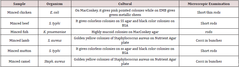
Table 1: Cultural Characteristic of isolates detected in meat samples.

Lupinepublishers-openaccess-complementary-alternative-medicine-journal

Result

In this study, different samples such as beef, fish, mutton, chicken, lamb and camel meat were analyzed which shows that meat sample is contaminated with foodborne pathogens such as E. coli, Staph. aureus, S. typhi, Klebsiella pneumoniae. It was found that 33% isolates of S. typhi and S. aureus were found whereas 17% isolates of E. coli and Klebsiella pneumoniae were detected from various meat samples. Most of the isolates were members of the Enterobacteriaceae so they differentiate based on lactose fermenter and nonlactose fermenter. E. coli and Klebsiella gave pink colored colonies on MacConkey agar while S. typhi gave colorless colonies. Biochemical testing was performed to differentiate among Enterobacteriaceae and identification of bacterial species. E. coli gave indole and methyl positive while Voges Proskauer and citrate negative. Klebsiella pneumoniae gave a positive VP test and citrate utilizing test.

In TSI test, acidic slope and acidic butt was found with gas production. S. typhi gave only MR test positive and in TSI it gave alkaline slop, acid butt with H2S production. All presumptively identified cultures were plated onto the selective medium, such as E. coli was inoculated on EMB agar. After incubation, it gives green metallic sheen. S. typhi was streaked on BSA and it gives black centered colonies with a black zone with metallic sheen surrounding the colonies. The positive result of catalase and coagulase test confirms the presence of Staph. aureus. All the identified isolates were susceptible to streptomycin, ceftriaxone, and sulfamethoxazole with average zones of inhibition at the respective concentrations as shown in Table 3 while staph. aureus was found resistant to oxacillin and s. typhi was found resistant to tetracycline.

Table 2: Antibiotic susceptibility testing.

Lupinepublishers-openaccess-complementary-alternative-medicine-journal

Discussion

This study was conducted to investigate the microbial load of foodborne pathogens in different raw meat sample which is available in open retail shops of Karachi. The presence of bacteria in raw meat has been widely reported from different parts of the world [24]. Some reports have recognized the presence of viable bacteria, especially gram-negative organisms from 106 to 109, as an indication of open-air meat spoilage [51]. In this study we detected the presence of pathogenic bacteria in raw meat including salmonella spp. (33%), Escherichia coli (17%), staphylococcus aureus (33%), and Klebsiella pneumonie (17%) which reflects the role of animal meat as a major reservoir for spreading pathogenic agents. Therefore, meat handlers and sellers should be made aware of the adverse effects of a lack of proper personal and environmental hygiene, along with sanitation.

Also, consumers should properly wash and cook the to avoid cross-contamination [25]. In this study raw chicken sample was found to be contaminated with E. coli (n=17). The presence of this indicator organism in raw meat might have originated from animal tissues or contaminated tools used during slaughtering and related treatment or cutting process. The identification of E. coli in this study showed the presence of fecal contamination and previous studies have shown that the presence of these organisms in food makes food unhealthy for consumption [26]. The high rate of prevalence of S. aureus (n=33) in raw lamb and camel meat indicates the presence of cross-contamination, which is usually related to human skin and clothing. This level of food contamination by this pathogen might lead to the food intoxications. Moreover, it is a major target in the screening of slaughterhouse carcasses to monitor hygienic conditions [27]. Raw beef and mutton sample were found to be contaminated with Salmonella typhi (n=33). The presence of S. typhi in raw meat indicates that the contamination is from human origin and may be the result of poor personal hygiene during the handling and processing of food. Salmonella continues to be a serious threat to consumer health not only due to its pathogenicity but also to its ability in adapting many different environments. The results of this study indicate that the rate of salmonella contamination in retail meat samples were high, ranging from 17% from beef and mutton samples [28]. In our study Klebsiella pneumonie (n=17) isolates were detected from the raw fish meat sample. A high incidence of Klebsiella spp from raw meat was also reported previously. Our data confirmed that the sold raw meat is of poor bacteriological quality and poses a high risk for consumer health. All the identified isolates were susceptible to streptomycin, ceftriaxone, and sulphamethoxazole antibiotic while S. typhi was found resistant against tetracycline. Staph. aureus showed resistance to oxacillin so it can be said that staph. aureus may have the ability to produce beta-lactamases enzyme.

In the developing world, foodborne infection leads to the death of many children, as well as resulting in diarrheal disease which can have long-term effects on children’s growth as well as on their physique To reduce the foodborne pathogens, like E. coli, staph. aureus Klebsiella and salmonella contamination rates in retail raw meats, it is critical that risk reduction strategies are used throughout the food chain. These strategies include on-farm practices that reduce pathogen carriage, increased hygiene at both slaughter and meat processing, continued implementation of HACCP systems, and increased consumer education efforts. Additionally, consumption of undercooked meat products and cross-contamination during food handling and preparation must be avoided to ensure food safety at home and in the foodservice industry.

For More Lupine Publishers Open Access Journals Please visit our Website:
To Know More About Open Access Publishers Please Click on Lupine Publishers

Follow on Twitter:  https://twitter.com/lupine_online

https://lupinepublishers.com/complementary-alternative-medicine-journal/fulltext/diversity-of-food-borne-pathogens-isolated-from-raw-meat-sample.ID.000141.php

Lupine Publishers | Cytoprotective Effect of Zingiber Officinale extract on Alcohol Induced Gastric Lesion in Mice

 Lupine Publishers | Open access Journal of Complimentary and Alternative Medicine


Ginger is a perennial plant that grows in India, China, Mexico and several other countries. The rhizome is used as both spice and in herbal medicine. Many studies have been done on pharmacological effect of ginger extract and our paper has been focused on cytoprotective effect of ginger on ethanol induced gastric injury in mice. Many drugs have been used in the treatment of peptic ulcer but each of them associated with different side effect so trends today toward use of herbal medicine which was associated with adverse effect. Ginger hydroalcoholic extract contain different consistent with anti-ulcer properties like 6-Gingersulfonic acid and three monoacyldigalactosyl glycerol including ginger glycolipid A. B. C .The mechanism of ginger extract may be due to counteracting the active oxidant radicals, decreasing mucosal cell shedding and thicking the mucus membrane so ginger extract has antiulcer properties when used in accurate dose and accurate duration.

Keywords: Cytoprotective effect; Ethanol; Ginger hydroalcoholic extract; Mice; Zingiber Officinale

Introduction

Spices, the predominant flavoring, coloring, and aromatic agents in food and beverages are now gaining importance for their diversified uses. Ginger (Zingiber Officinale) is a medicinal plant that has been widely used in Chinese, and Tibb Unani herbal medicines all over the world, since antiquity, for a wide array of unrelated ailments that include muscular aches, sore throat, constipation, arthritis, indigestion, vomiting and infectious diseases. Currently, there is a renewed interest in ginger, and several scientific investigations aimed at isolation and identification of active constituents of ginger, scientific verification of its pharmacological actions and of its constituents, and verification of the basis of the use of ginger in several diseases and conditions Ali et al. [1].

Ginger grows best in tropical and subtropical areas, which have good rainfall with hot and humid conditions during the summer season. It is a member of Zingiberaceae family originated in Southeast Asia and has been introduced to many parts of the globe where it proliferates in suitable environment. Belief in the medicinal properties of ginger existed in ancient Indian and oriental cultures where ginger has been used alone or as a component in herbal remedies. This practice continues today in many areas of the world including Africa, Brazil, China and, Mexico. Ginger has introduced to Europe and other areas by Dutch, Portuguese Arab and Spanish explorers or traders from around the 13th to 16th centuries.

Carbohydrates

Starch is the major constituent up to 50%.

Oleoresin

Gingerol homologues (major, about 33%) include derivatives with methyl side chain, shogaol homologues (dehydration products of gingerols), zingerone (degradation product of gingerols) ,1- dehydrogingerdione and 6- ginger sulfonic acid.

Lipids 6-8%

They include free fatty acids e.g. palmitic acid, oleic acid, linoleic acid, caprylic acid, capric acid, lauric acid, myristic acid, pentadecanoic acid, heptadecanoic acid, stearic acid, linolenic acid, arachidonic acid, triglycerides, phosphatidic acid, lecithin and ginger glycolipids A, B and C. Ulcer are caused due to imbalance between aggressive factors (hydrochloric acid, pepsin, gastrin, no steroidal anti-inflammatory drugs, and ethanol) and defensive factor of gastric mucosa (prostaglandin, mucus, bicarbonate). The antiulcerogenic activity of many plant products is reported due to an increase in mucosal defensive factors rather than decrease in the aggressive factors Goel, et al. [2]. Numbers of an antiulcer drugs like gastric antisecretory drugs H2- receptor antagonist, antimuscarinic agents, proton pump inhibitors, and mucosal protective agents, carbenoxolone sodium, sucralfate, and prostaglandin analogous are available which are shown to have side effects and limitation Baowman et al. [3].

There are several herbal ayuvedic preparation which have a protective effect against gastric mucosal injury Shetty et al. [4]. Herbal medicine is now used by up to 50% of the Western population in a number of instances 10% for treatment or prevention of digestive disorders Langmead and Rampton [5]. Today, pharmacopoeias of a number of different countries list ginger extract for various digestive disease, Aromatic, spasmolytic and carminative properties of ginger are probably responsible for the therapeutic application in digestive tract ailments Sertie et al. [6].

Mechanism of Gastric Effect of Ginger

Several studies have shown that ginger extract, essential oils and glycolipids possess a number of pharmacological actions, which at least in part for some of them anti-ulcerogenic or ulcer preventive efficacy may be suggested. Common side effects of treating inflammation with modern drugs is that ulcer in the digestive system can be created or their condition made worse Wallace [7]. Ginger not only relieves the symptoms of inflammation but also protects the creation of digestive ulcers. Ginger may protect the stomach from the damaging effect of alcohol and non- steroidal anti-inflammatory drugs and may help prevent ulcers Villegas et al. [8]. A study was done in Isfahan university by Miniaiyan et al. [9] for anti-ulcerogenic effect of ginger on cysteamine-induced duodenal ulcer in rats. The results obtained in positive control groups indicated that ginger possesses its anti-ulcerative properties through a mechanism mainly related to acid- pepsin inhibition.

The effect of ginger (acetone extract) and zingiberene on hydrochlochloric acid/ethanol-induced gastric lesions in rats have been examined (6)- gingerol and zingiberene, both 100mg/ kg oral), significantly inhibited gastric lesions by 54.5% and 53.6% respectively. The total extract inhibited lesions by 97.5% at 1g/kg. Oral administration of both aqueous and methanol ginger extract to rabbit has been reported to reduce gastric secretions (gastric juice volume, acid and pepsin output). Both extracts were found to be comparable with cimetidine (50mg/kg) with respect to gastric juice volume.

The aqueous extract was comparable with cimetidine and superior to the methanol extract for pepsin output, and the methanol extract superior to the aqueous extract and comparable to cimetidine for acid output. Roasted ginger decoction which showed an obvious inhibiting tendency on three gastric ulcer models. The plant contains active materials which for some of them ulcer protective properties have been identified 6- ginger sulfonic acid and three monoacyldigalactosyl glycerols including ginger glycolipid A,B,C have been isolated from dried rhizome of Zingiber Officinale which are potent anti-ulcer components Yoshikawa et al. [10].

a) 6- gingerol and 6- shagaol are two other anti-ulcer components that are less potent nut are mainly responsible for ginger pungency.

b) Results of one study fore effects of three herbal medicine including Zingiber Officinale on gastric ulceration and secretion in rats indicated significant protection against stress, aspirin and pylorus ligation. The proposed anti-ulcerogenic effects were augmentation of mucin secretion and decrease in cell shedding.

c) Al-Yahya et al. [11] Studied the cytoprotective and gastric anti-ulcer effect of ginger in albino rats. Cyto destruction was produced by 80% ethanol, 0.6M HCL, 0.2 M NaOH and 25% Nacl. Gastric ulcer was produced by ulcerogenic agents including indomethacin, aspirin and reserpine beside hypothermic restraint stress and by pylorus ligation. The results of this study demonstrated that the extract in the dose 500mg/kg orally exerted highly significant cryoprotection and prevented occurrence of gastric ulcers induced by non- steroidal antiinflammatory drugs.

Method

Experimental animals: Albino mice of either sex weighing 20- 30g, was maintained in the animal house of Faculty of Medicine- Al Arab Medical University, Benghazi, Libya the mice were bred in the faculty animal house. All animals were housed in standard polypropylene cages (48×35×22cm) and kept under controlled room temperature. (20±5 °C; relative humidity 60-70%) in a 12h light-dark cycle.

The animals were given a standard laboratory diet and free water Food was withdrawn 12h before and during the experimental hours:

Maceration method

In this method fresh ginger rhizome was cut into small pieces, dried, and then pulverized into coarse powder and weighing about 400g of powder. It was macerated in 1000ml hydroalcoholic solution (70% Ethanol, 30% distilled water) for seventy-two hours. The extract was then shaked, filtered by using dryness filter paper and the solution was evaporated in a rotatory evaporator under reduced pressure until evaporation and removal of the solvent give hydroalcoholic extract of ginger out of 400g of crude plant, 8g of hydroalcoholic extract of ginger were obtained and kept for use in pharmacological experiments (Iranian Herbal Pharmacopeia). Study of cytoprotective effect of Zingiber (ginger) extract on ethanol-induced gastric lesion in mice.

The present paper was designed to investigate the cytoprotective effect of hydroalcoholic extract of ginger in an animal model of gastric lesion. Mice of either sex weighing 25-30g were employed in this study the animals were divided isoups each consisting of six mice.

1. Normal group: Given vehicle (5ml/kg orally) without ulcer induction.

2. Control group: Given vehicle (5ml/kg orally) one hour before ulcer induction.

3. Extract group: Given hydroalcoholic extract of ginger in a dose (300mg/kg orally) one hour before ulcer induction.

4. Extract group: Given hydroalcoholic extract of ginger in a dose of (600mg/kg I.P) one hour before ulcer induction.

5. Chronic extract group: Given hydroalcoholic extract of ginger. In a dose of (300mg/kg I.P) for five consecutive days before ulcer induction. The last dose was administered one hour before ulcer induction.

6. Reference group: Given rantidine (50mg/kg I.P) one hour before ulcer induction.

One hour after intragastric administration of absolute ethanol (95%, 0.2ml), the animals are euthanized with ether, the stomach were excised, cut along the greater curvature, and gently rinsed under tap water, and examined by 5-fold binocular magnifier to assess lesion in gastric mucosa.

Results

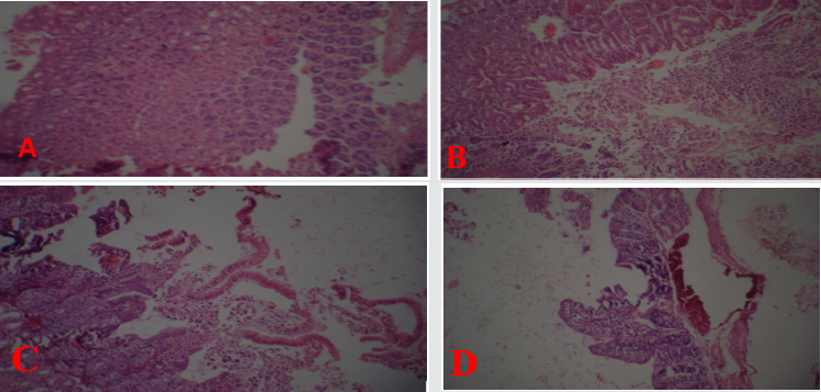
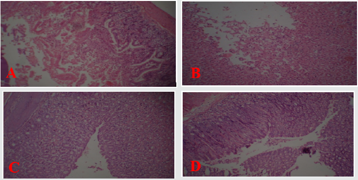
As shown in Figure 1 normal gastric mucosa Figure [A] compared with Figure [B & C] with treatment by ethanol there is exfoliation and sloughing of gastric cells with inflammatory cell infiltrate and congestion of blood vessels Figure [D]. Figure 2 shows partial improvement in gastric lesion in the group treated by oral ginger treatment at dose 300mg/kg as there is mild exfoliation of gastric cells Figure [A]. Moderate gastric protection by (I.P) treatment of ginger at a dose 600mg/kg as there is moderate exfoliation of gastric cells as shown in Figure [B]. Complete protection by chronic (I.P) treatment of ginger in a dose of 300mg/kg with no exfoliation of gastric cells as shown in Figure [C]. Rantidine has moderate protective effect as shown in Figure [D] compared with extract in Figure [C] with complete protection of gastric mucosa.

Figure 1: Light photomicrographs (10× magnifications) of hematoxylin and eosin- stained sections of stomach.

A: Normal gastric mucosa.

B, C: Ethanol treated stomach with Exfoliation and sloughing of gastric cells.

D: Ethanol treated with congestion of blood vessel.

Lupinepublishers-openaccess-complementary-alternative-medicine-journal

Figure 2: Light photomicrographs(10×magnifications) of hematoxylin and eosin-stained sections of stomach:

A: Ginger treated (IP) stomach.

B: Ginger treated (IP) stomach.

C: Chronic ginger treated (IP).

D: Ranitidine treated (IP) stomach.

Lupinepublishers-openaccess-complementary-alternative-medicine-journal

In present paper ethanol administration provoked significant gastric mucosal injuries as evidence by necrosis, exfoliation and sloughing of gastric cells, congestion of blood vessels and infiltration of inflammatory cells. Cytoprotective action of ginger has been investigated using animal models of acute gastric injury induced by necrotizing agents such as ethanol. Ethanol serves as the most common ulcerogenic agent and produced sever gastric hemorrhagic erosions Robert et al. [12]; Szabo et al. [13]. The genesis of ethanol-induced gastric lesions is multifactorial with the depletion of gastric wall mucus content as one of the involved factors. Submucosal venular constriction by ethanol and eventual injury is caused due to perturbations of superficial mucosal cells, notably the mucosal mast cells leading to release of vasoactive mediators including histamine, that cause damage to gastric mucosa Hollander et al. [14]. Accumulation of activated neutrophil in gastric mucosa may be the source of free radicals Oxygen free radical which lead to increase to lipid peroxidation and damage to cell membrane are impacted in ethanol induced gastric mucosal injury Al-Harbi et al. [15]. In addition to its direct damage of gastric mucosal cells by development of free radicals, cause solubilization of mucus constituents and depressant tissue levels of protein leading to flow stasis in gastric blood. The present paper revealed parial cytoprotection by single oral dose of ginger (300mg/kg) in mice. More effective cytoprotection was elicited by (600mg/kg I.P).

The results obtained in the present paper indicated that ginger extract possess its anti- ulcerative properties through a mechanism mainly related to acid and pepsin inhibition Chronic I.P injection of plant extract in a dose of (300mg/kg) more effective in prevention lesion formation. The mechanism of ginger extract in chronic (I.P) protection may be due to counteracting the active oxidant radicals, decreasing mucosal cell shedding and thicking the mucus membrane. The mechanism of anti-ulcer effect of ginger may be due to presence of 6-Gingersulfonic acid and three monoacyldigalactosyl gylcerols including ginger glycolipid A. B. C have been isolated from dried rhizome of Zingiber Officinale which are potent anti-ulcer components Yoshikawa et al.[16]. 6- Gingerol and 6- shogaol are two other anti- ulcer components that are less potent but are mainly responsible for ginger pungency. The results of present paper in accordance with previous reports in which water and methanolic extract of eight Zingiberaceae herbs caused a significant decrease in gastric secretion in un-anesthetized rabbits and the effect of water extract was similar to cimetidine Sakai et al. [17].


For More Lupine Publishers Open Access Journals Please visit our Website: https://lupinepublishers.us/
To Know More About Open Access Publishers Please Click on Lupine Publishers

Follow on Twitter:  https://twitter.com/lupine_online

https://lupinepublishers.com/complementary-alternative-medicine-journal/fulltext/cytoprotective-effect-of-zingiber-officinale-extract-on-alcohol-induced-gastric-lesion-in-mice.ID.000139.php

Lupine Publishers | Tongluo Decoction Blocks Apoptotic Mitochondrial Pathway and Inhibits Cardiomyocyte Apoptosis of Coronary Microembolization Rats

 Lupine Publishers| Open access Journal of Complimentary and Alternative Medicine 


BackgrBackground: We found that Tongluo Decoction has had good protection for myocardial cells injury of CME rats. But it is not clear whether Tongluo Decoction has inhibitory effect on myocardial apoptosis. So we want to study the effect and mechanism of Tongluo decoction to cardiomyocyte apoptosis following coronary microembolization in rats. Methods: 46 male Wistar rats were randomly divided into blank group (n=6), model group (n=10), Tongluo Decoction low dose group (n=10), Tongluo Decoction high dose group (n=10) and Tongxinluo group (n=10). In addition to the blank group, the rats of other groups were established the model of autologous thromboembolism microembolism by interventional method. The low dose group of Tongluo Decoction was given 7.8g/kg for gavaging, the high dose group of Tongluo Decoction was given 23.4g/kg, Tongxinluo group was given 0.2g/kg, once-a-day, for 12 day. The judgment of the model was done with HE and HBFP staining, the myocardial apoptosis rate was observed by TUNEL staining. Western Blot was used to detect Bcl-2, Bax and Caspase-3 in cardiac myocytes.

Results: The microembolization vessel count ratio, ischemic myocardial area ratio and the apoptosis rate of myocardial cells in model group were significantly higher than that in blank group, bcl-2 and bcl-2/bax decreased, and bax and caspase-3 were significantly increased (P < 0.05).Compared with model group, Tongluo Decoction low dose group, Tongluo Decoction high dose group and Tongxinluo group microembolization vessel count ratioischemic myocardial area ratio apoptosis rate decreased (P < 0.05), bcl-2, bcl-2/bax increased, Bax and caspase-3 decreased (P < 0.05). Compared with Tongxinluo group, the ratio of ischemic myocardial area, microembolization vessel count ratio, apoptosis rate, the expression of bax and caspase-3 proteins decreased (P > 0.05), bcl-2 and bcl-2/bax ratio increased in Tongluo Decoction high dose group (P > 0.05). bcl-2 and bcl-2/bax ratio increased obviously in Tongluo Decoction low dose group (P < 0.05).

Conclusion: Tongluo Decoction can inhibit cardiomyocyte apoptosis following coronary microembolization in rats, and its possible mechanism is related to blocking mitochondrial apoptotic pathway.

Keywords: Tongluodecoction; Rat; Coronary microembolization; Cardiomyocyte apoptosis

Abbrivations: Percutaneous coronary intervention (PCI); Coronary microembolization (CME); Hematoxylin-Eosin staining (HE); Hematoxylin basic fuchsin picric staining (HBFP); Terminal deoxynucleotidyl transferase-mediated dUTP Nick End-Labeling (TUNEL); High performance liquid chromatography (HPLC); Tongluo decoction (TLD); Optical density (OD); Bcl-2 Assaciated X protein (Bax); Cysteinyl aspartate specific protease-3 (Caspase-3); B-cell lymphoma-2 (Bcl-2)

Introduction

In the process of percutaneous coronary intervention which is used to treat coronary heart disease, the small fragments that come from mechanical injury of coronary atherosclerosis, with the flow of blood circulation to the distal coronary artery, block the distal microvascular, which leading to the obstruction of coronary microvascular circulation, that is coronary microembolization(CME). CME is a serious complication of PCI, which can cause myocardial ischemia and cardiac dysfunction, and even lead to death. Numerous experimental studies [1, 2] have shown that apoptosis plays an important role in CME lesions. The research showed that Chinese materia medica can improve myocardial injury caused by coronary microembolization [3]. We found that Tongluo Decoction has had good protection for myocardial cells injury of CME rats. The Tongluo decoction could inhibit the pathological change of CME in rats, reducing the ingury of myocardial cells, inhibiting the expression of inflammatory cytokines and promoting the microemboli vascular recanalization [4]. But it is not clear whether Tongluo Decoction has inhibitory effect on myocardial apoptosis of CME rats. Therefore, in this study, we established coronary artery microembolization model of rats by injecting autologous thrombus through the right common carotid artery by interventional method with balloon catheter [5]. The effect and mechanism of Tongluo Decoction on cardiomyocyte apoptosis following CME in rats were studied.

Methods

Preparation and quality control of tongluo decoction

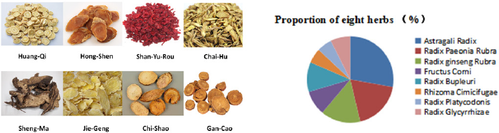
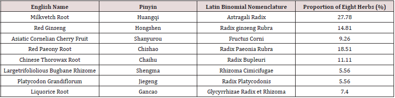
Tongluo Decoction mainly consisted of eight herbs: [6- 8] The composition and proportion of herbs are shown in the diagram (Figure 1, Table 1). The herbs was purchased from the Hui ren Tang Pharmaceutical Company, and they were identified by Jianyin Li, a teacher of Lanzhou University school of pharmacy.(The voucher specimen numbers of the stored herbarium specimen are 62032120180807063LY for Astragali Radix, 620031220010918321LY for Radix ginseng Rubra, 62062319980910435LY for Fructus Corni, 62032120180811156LY for Radix Paeonia Rubra, 62032120180807062LY for Radix Bupleuri, 62062320130815322LY for Rhizoma Cimicifugae, 62032120180805049LY for Radix Platycodonis, 62032120180812164LY for Radix Glycyrrhizae. ) This herbs that was conformed to the prescription, all of them were qualified, decocted, concentrated, and then put in refrigerator at 4. The chromatographic conditions were Diamonsil C18 (2) column (200×4.6mm, 5μm), the liquid phase of the calycosin glycoside (batch number 111920-201606, National institutes for food and drug control) mobile phase: acetonitrile -0.2% formic acid solution (15:85), flow velocity:1ml/min, column temperature: 30, detection wavelength: 260nm, sample size: 10μl. Paeoniflorin (batch number X27F8C30162, Shanghai source leaf Biological Technology Co., Ltd.) mobile phase: acetonitrile-0.1% phosphoric acid solution (16:84). Flow velocity, column temperature and sample size remain unchanged. Detection wavelength: 230nm; Ginsenoside Rb1 (batch number Z06M8L30693, Shanghai source leaf Biotechnology Co., Ltd.) mobile phase: acetonitrile -0.1% phosphoric acid solution, gradient elution. Flow velocity, column temperature and sample size remain unchanged. The detection wavelength: 203nm. To calculate the content of calycosin glycoside, paeoniflorin and ginsenoside Rb1.

Figure 1: Composition of main herbs and their proportion in Tongluo Decoction.
Lupinepublishers-openaccess-complementary-alternative-medicine-journal

Table 1: The full scientific species names of ingredients of Tongluo Decoction.
Lupinepublishers-openaccess-complementary-alternative-medicine-journal

Preparation of autologous thrombus suspension

1ml blood was collected from the caudal vein of each rat , then drying, the dry blood was grinded into powder like particles. The thrombus particles with a diameter between 41μm and 80μm were mixed into a suspension, and 2×10⁵ thrombus particles were injected into the the aortic root through the right common carotid artery for establishing coronary artery microembolization model.

Animals grouping and model establishment

46 SPF healthy adult male Wistar rats (weight 350g±30g) were randomly divided into blank group (n=6), model group (n=10), Tongluo Decoction low dose group (n=10), Tongluo Decoction high dose group (n=10), Tongxinluo group (n=10). The experimental animals were purchased from the experimental animal center of Lanzhou University. The laboratory animal license number SCXK (Gan) 2013-0002. The experimental process adhered to the ethical regulations of animal experiments. The rats were anesthetized with 0.4% pentobarbital sodium solution, separated the right common carotid artery and cut the “V” incision at its distal end. The balloon catheter, which was soaked with heparin sodium saline, was pushed to the root of the aorta (about 4cm from the incision of right common carotid artery to the aortic root ), and the blood flow of the ascending aorta was blocked for 3-5s, rapidly withdrawing the guide wire during the period, and the autologous thrombus suspension was injected. Balloon catheter was pulled out, the proximal heart segment of the right common carotid artery was ligated. Penicillin 4×10⁵ U/each was given for 3 days after the operation to prevent infection.

Drug intervention

The daily dosage of the low dose group of Tongluo Decoction was calculated according to the conversion ratio of the 1:6.25 in the rats [5]. The dosage of the high dose group of Tongluo Decoction was 3 times that of the low dose group of Tongluo Decoction. Tongxinluo capsule (Shijiazhuang Yiling pharmaceutical Limited by Share Ltd) uses 9grains/day dose, dissolve it in double steamed water and intragastric gavage. Tongxinluo capsule is purchased from Hui Ren Tang Pharmaceutical Company and its specification is 0.26g/ granules. The rats of each group were given the corresponding drug by intragastric gavage for 12 days. During the test, 3 rats died in each of the model group, low dose group of Tongluo Decoction, high dose group of Tongluo Decoction and Tongxinluo group during the modeling process, and 1 died in Tongxinluo group after gavage.

Collection of heart specimens

After the drug intervention, all rats were sacrificed by cervical dislocation under anesthesia by intraperitoneally injected 0.4% sodium pentobarbital solution 1ml per 100g body weight. Then rat heart was collected. The apex of heart was fixed in 4% polycondensation formaldehyde solution. After dehydration and paraffin embedded, 5μm consecutive paraffin sections were used to HE and other staining. The base of the heart was immediately placed in a pre-freezing storage tube, stored at liquid nitrogen, and used to detect cardiomyocyte apoptosis protein by Western Blot.

HE and HBFP staining of rat myocardium

After the apex staining of heart paraffin section of rats in each group with HE and HBFP, the staining sections were observed by three researchers under 400× and 100× microscope respectively to calculate the microembolization vessel count ratio, ischemic myocardial area ratio.

Detection of cardiomyocyte apoptosis by TUNEL

The pathological tissue section of the rat in each group was treated with anti-shedding. According to the TUNEL instructions (Boster Co., Ltd., Wuhan) to stain. The number of cardiomyocyte apoptosis and the number of total cells under the microscope were observed by three researchers under 400× microscope, and the apoptosis rate of myocardial cells was calculated and the apoptosis level of cardiac myocytes was evaluated. Apoptosis rate (%)=(the number of apoptotic cells/the total cell number under microscope)×100.

Western Blot was used to detect the expression of cardiomyocyte apopt-osis proteins

To extract the protein of myocardial tissue, according to the instructions of the kit (Solarbio Science and Technology Co., Ltd. Beijing). The OD value was measured, and the standard protein curve was drawn. Then calculating the protein concentration. Prepare 12% separation glue and 5% concentration glue, 160 V constant pressure electrophoresis 100min, and PVDF membrane was then transferred, followed by incubation for 2h with blocking buffer containing 5% Non-fat milk powder. Diluting and putting relevant primary antibodies, bcl2, bax, caspase3, and GAPDH respectively (Biosynthesis biotechnology Co., Ltd. Beijing), with gentle shaking overnight at 4. Afterwards, the membrane was incubated with secondary antibody(purchased from SAB) for 1h at room temperature, then washing and exposing.

Data statistics method

The SPSS 22.0 statistics software was used to analyze the data, and the measurement data were expressed with mean±standard deviation (x±s). Single factor variance( one-way ANOVA) was used in comparison of multiple groups. And P < 0.05 indicated that the difference was statistically significant.

Results

Quality control results of tongluo decoction

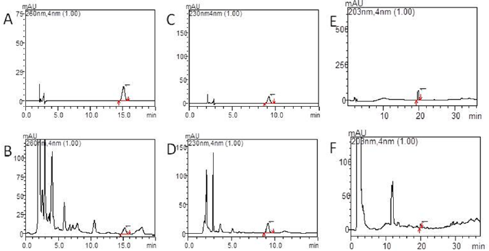
As shown in Figures 2(a)-2(f), the separation of the components to be tested is good and has no interference with other components. High performance liquid chromatography (HPLC) shows the peak appearance time of the calycosin glycoside standard and Tongluo Decoction for test sample was 15.0±0.5 min, while the paeoniflorin standard and Tongluo Decoction for test sample was 9.0±0.5 min, and the ginsenoside Rb1 standard and Tongluo Decoction was 20.0±0.5 min, and the effective components of three herbs are compared with their respective standard products, some impurity peaks appeared. However, the separation efficiency of the effective components of the three herbs were not affected. The above results indicated that the effective components of Tongluo Decoction contained of calycosin glycoside, paeoniflorin and ginsenoside Rb1. The results showed that the effective components of Tongluo Decoction included calycosin-7-glucoside, paeoniflorin and ginsenoside Rb1, and the quality of the decoction was stable and could be used for the follow-up drug intervention test.

Figure 2:

a) High performance liquid chromatography of Tongluo Decoction, calycosin-7-glucoside standard.

b) High performance liquid chromatography of Tongluo Decoction. A calycosin-7-glucoside standard, Tongluo Decoction, calycosin-7-glucoside.

c) High performance liquid chromatography of Tongluo Decoction,paeoniflorin standard.

d) High performance liquid chromatography of Tongluo Decoction, Tongluo Decoction, 1 is paeoniflorin.

e) High performance liquid chromatography of Tongluo Decoction, ginsenoside Rb1 standard.

f) High performance liquid chromatography of Tongluo Decoction, Tongluo Decoction, 1 is ginsenoside Rb1.
Lupinepublishers-openaccess-complementary-alternative-medicine-journal

HE staining results of rat myocardium

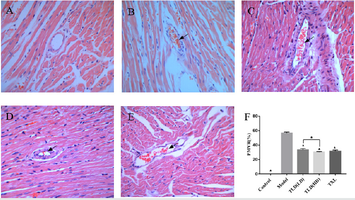
As shown in Figures 3(a)-3(f), the microembolus were not found in the microvessels of the coronary artery in the blank group. A large number of microembolus were found in the coronary microvessels in the model group, and around the microembolus there were inflammatory cell aggregation. Some of microembolus were found on the low dose group of Tongluo Decoction, the high dose group of Tongluo Decoction, and the Tongxinluo group in the coronary microvessel. The positive microembolization vessels count ratio (PMVR) in the model group was significantly higher than that in the control group (P < 0.05). Compared with the model group, the low dose group of Tongluo Decoction, the high dose group of Tongluo Decoction and the Tongxinluo group were significantly lower than those in the model group (P < 0.05). Compared with the Tongxinluo group, the PMVR in the Tongluo Decoction low dose group was increased while the Tongluo Decoction high dose group was decreased(P > 0.05). The PMVR of high dose group of Tongluo Decoction was lower than that in the low dose group of Tongluo Decoction (P < 0.05). (Figures 4(a)-4(f)).

Figure 3: Comparison of HE staining of myocardial positive microembolic blood vessels of rats in each group(400×). A blank group, B model group, C TLD(LD) group, D TLD(HD) group, E TXL group. F Comparison of HE staining of myocardial positive microembolic blood vessels of rats in each group. (▲)P<0.05, versus model group; (★)P<0.05, TLD(LD) group versus TLD(HD) group. PMVR:Positive microembolization vessel count ratio.
Lupinepublishers-openaccess-complementary-alternative-medicine-journal

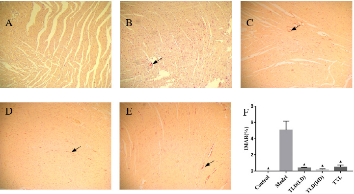
Figure 4: Comparison of HBFP staining of ischemic myocardium of rats in each group(100×). A blank group, B model group, C TLD(LD) group, D TLD(HD) group, E TXL group, F Comparison of HBFP staining of ischemic myocardium of rats in each group. (▲)P<0.05, versus model group. IMAR:Ischemic myocardial area ratio.
Lupinepublishers-openaccess-complementary-alternative-medicine-journal

HBFP staining results of ischemic myocardium

In Figures 5(a)-5(f), almost no red myocardium tissues was found in the blank group. A large number of red myocardium tissues were found in the model group. Tongluo Decoction low dose group, Tongluo Decoction high dose group and Tongxinluo group found some red myocardium tissues. Compared with the blank group, the ratio of ischemic myocardial area ratio in the model group was significantly higher (P < 0.05). Compared with the model group, the ratio of ischemic myocardial area ratio in Tongluo Decoction low dose group, Tongluo Decoction high dose group and Tongxinluo group decreased significantly (P < 0.05). Compared with Tongxinluo group, the ratio of ischemic myocardial area ratio(IMAR) in Tongluo Decoction low dose group, Tongluo Decoction high dose group was decreased, but the difference was not statistically significant (P > 0.05) .

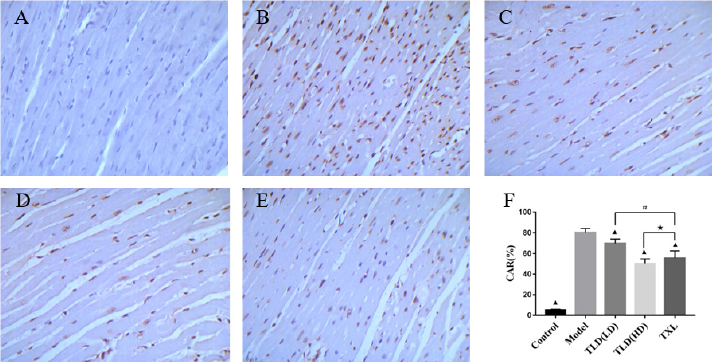
Figure 5: Comparison of TUNEL staining(400×)and apoptosis rate of myocardial cells in each group of rats. A blank group, B model group, C TLD(LD) group, D TLD(HD) group, E TXL group. F Apoptosis rates of cardiomyocytes in TUNEL staining groups were compared, (▲)P<0.05, versus model group; (#)P<0.05, TLD(LD) group versus TXL group; (★)P>0.05, TLD(HD) group versus TXL group. CAR: cardiomyocyte apoptosis ratio.
Lupinepublishers-openaccess-complementary-alternative-medicine-journal

Detection of cardiomyocyte apoptosis by TUNEL

As shown in Figure 6, TUNEL staining showed that the normal nuclei were pale blue, the apoptotic nuclei were tan, nuclear chromatin was concentrated, and the nuclei were broken into unequal nucleus fragments, accompanied by the formation of apoptotic bodies. There were almost no tan nuclei in the control group, and a number of tan nuclei in the model group. Tongluo Decoction low dose group, Tongluo Decoction high dose group and Tongxinluo group have some tan nuclei. The apoptosis rate of myocardial cells in the model group was significantly higher than that in the control group (P < 0.05). Compared with the model group, the cardiomyocyte apoptosis rate in Tongluo Decoction low dose group, Tongluo Decoction high dose group and Tongxinluo group decreased significantly (P < 0.05). Compared with Tongxinluo group, the decrease of cardiomyocyte apoptosis rate in Tongluo Decoction low dose group was small (P < 0.05) , but the decrease of cardiomyocyte apoptosis rate in Tongluo Decoction high dose group was much more (P > 0.05).

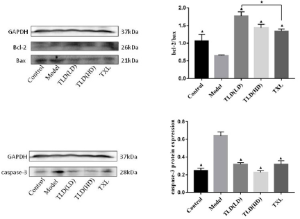
Figure 6: Western Blot detection of bcl-2、bax、caspase-3 protein expression in cardiac myocytes.
Lupinepublishers-openaccess-complementary-alternative-medicine-journal

Western Blot detect the expression of Bcl-2, Bax and caspase-3

Protein in Cardiac Myocytes

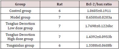
As shown in Figures 3(a)-3(f)) and (Table 2), compared with the control group, the expression of Bcl-2 protein and bcl-2/bax of the model group were decreased, the expression of Bax and Caspase-3 in the model group were significantly increased (P < 0.05). Compared with the model group, the expression of Bcl-2 protein, bcl-2/bax were increased and the expression of Bax and Caspase-3 were decreased in the Tongluo Decoction low dose group, Tongluo Decoction high dose group and Tongxinluo group (P < 0.05). Compared with Tongxinluo group, the expression of Bcl-2 protein, bcl-2/bax increased (P < 0.05), the expression of Bax and caspase-3 protein decreased in the Tongluo Decoction low dose group and the Tongluo Decoction high dose group (P > 0.05). These results indicate that the coronary microembolization(CME) can induce cardiomyocyte apoptosis, and Tongluo Decoction and Tongxinluo can inhibit cardiomyocyte apoptosis.

Table 2: The results of bcl-2/bax ratio in each group of rats (x ± s ).
Lupinepublishers-openaccess-complementary-alternative-medicine-journal

Note: Comparison with control group.

aP﹤0.05; Comparing with model group.

bP﹤0.05; Comparing with Tongxinluo group, cP﹤0.05.

Discussion

CME is an common complication in the interventional therapy of coronary heart disease. It is one of the important reasons for the occurrence of “no reflow” and “slow flow” of coronary artery. It is an important factor of poor prognosis and even death in some patients after PCI. The secondary myocardial apoptosis and necrotic myocardium irreversible injury are independent predictors of no reflow and poor prognosis. Research shows [4] that a stable, reliable and economical model of coronary arteria microembolism can be established by injecting autologous thrombus to the aortic root of rat through the right common carotid artery. In this study, we followed this method and established the rat CME model successfully. After the drug intervention 12 days, we found that both Tongluo Decoction and Tongxinluo could contribute to the narrowing of the microembolus, promote the blood circulation of the coronary microembolic vessels and improve the condition of myocardial ischemia. Tongluo Decoction is composed of many kinds of Chinese medicinal materials according to the rule of monarch(Jun), minister(Chen), assistant(Zuo) and guide(Shi). It has the effects of benefiting qi for activating blood circulation and disperse blood stasis and dredge collateral circulation. Among this Chinese medicine compound, Astragali Radix(Huangqi) as the monarch, compatible with Radix ginseng Rubra (Hongshen) can tonify Qi of spleen and Lung significantly. It made QI and blood own a recourse to metaplasia. Compatibility of Radix Paeonia Rubra (Chishao) have an effect on blood stasis runs and relieve pain, cooling blood and subsidence of a swelling. Assistance of Radix Bupleuri(Chaihu), Rhizoma Cimicifugae(Shengma), Radix Platycodonis(Jiegeng) for lifting Yangqi and carrying medicine upgoing. Qi running leads to blood circulation. Then add Fructus Corni(Shanyurou) to nourish the Yin of liver and kidney. Radix Glycyrrhizae(Gancao) as guide plays an harmonization of all drugs role in the compound. All drug compatibility, reflect a efficacy of tonifying qi and yin and promoting blood circulation to remove meridian obstruction. Zhang Zhenguo and other studies [6] found that Tongluo Decoction could inhibit the pathological process of coronary microembolism, reduce myocardial damage, inhibit the expression of inflammatory factors and promote the role of microembolic vascular recanalization.

Apoptosis is an active and orderly death process, which is produced by the body in the physiological and pathological conditions to maintain the homeostasis of its own internal environment [7]. Studies have shown that apoptosis is one of the possible mechanisms of ventricular dysfunction induced by coronary microembolization [8]. Anti apoptotic therapy can significantly inhibit the apoptosis of cardiomyocytes after CME in rats [9-11]. Studys showed that Tongxinluo capsule could inhibit cardiomyocyte apoptosis [12, 13]. In our study, Tongxinluo was used as a positive control group to further study the mechanism of Tongluo Decoction from the perspective of apoptosis. Mitochondria mediated apoptosis pathway is one of the most important apoptotic pathways. Numerous studies have shown [14-16] that inhibition of mitochondrial mediated apoptosis plays an important role in improving CME induced myocardial damage and cardiac dysfunction. The regulation of mitochondrial apoptosis is mainly mediated by the regulation of Bcl-2 family proteins [17, 18]. After the destruction of mitochondria, the release of cytochrome C increased. Under the regulation of Bcl-2 family protein, the release of cytochrome C was induced by anti apoptotic members and inducing apoptotic members, eventually causing the activation of Caspase-3, resulting in the apoptosis [19]. Cysteinyl aspartate specific protease (caspase) is the main effector factor of apoptosis.

Wang JiangYou and other studies [20] showed that increased the expression of Caspase-3 in rat cardiomyocytes after CME, and pretreatment with atorvastatin could downregulate the expression of Caspase-3. Liu Tao and other studies [21] found that after CME bcl-2 expression decreased, Bax and Caspase-3 expression increased, silence LOX-1 can reverse Bcl-2, Bax, caspase-3 protein expression. Bcl-2 can inhibit apoptosis, while Bax can promote apoptosis [22]. The ratio of Bcl-2/Bax is an important parameter of mitochondrial signaling pathway, and the ratio between them is one of the important causes of apoptosis [23, 24] Our study showed that the apoptosis rate of myocardial cells in the CME model group was significantly increased. The expression of Bcl-2 protein, bcl-2/bax were decreased and the expression of Bax and Caspase-3 were significantly increased in the model group. It was indicated that CME induced the cardiomyocytes apoptosis. The apoptosis rate of cardiac myocytes in Tongluo Decoction low dose group, Tongluo Decoction high dose group and Tongxinluo group was significantly lower than that of model group (P < 0.05), and the expression of Bcl-2 protein and bcl-2/bax increased, and the expression of Bax and caspase-3 protein decreased (P < 0.05). It indicates that Tongluo Decoction and Tongxinluo capsule can inhibit the cardiomyocytes apoptosis of rat after CME. Compared with Tongxinluo group, the degree of decrease on cardiomyocyte apoptosis rate in Tongluo Decoction low dose group was lower (P < 0.05) while Tongluo Decoction high dose group was higher (P > 0.05). The results of Western Blot showed that, compared with Tongxinluo group, the expression of Bcl-2 protein, bcl-2/bax increased (P < 0.05), the expression of Bax and caspase-3 protein decreased in the Tongluo Decoction low dose group and the Tongluo Decoction high dose group (P > 0.05). It shows that Tongluo Decoction is almost equivalent to Tongxinluo capsule in the treatment of CME, which can inhibit the apoptosis of myocardial cells of rats after CME, and its mechanism may be related to the blocking of mitochondrial apoptosis pathway.

Conclusion

Tongluo Decoction may inhibit the cardiomyocytes apoptosis of CME rats by blocking mitochondrial apoptosis pathway and protect their myocardium.

For More Lupine Publishers Open Access Journals Please visit our Website:
To Know More About Open Access Publishers Please Click on Lupine Publishers

Follow on Twitter:  https://twitter.com/lupine_online

https://lupinepublishers.com/complementary-alternative-medicine-journal/fulltext/tongluo-decoction-blocks-apoptotic-mitochondrial-pathway-and-inhibits-cardiomyocyte.ID.000138.php

Friday, February 21, 2020

Lupine Publishers | Is There Any Relation Between Blood in Urine and Eating Almonds?


Lupine Publishers | Open access journal of Complimentary & Alternative Medicine


Abstract


The main aim of the study is to find out the relation of eating almond with the blood in urine. This is the alarming situation when a person sees blood in the urine, it is called hematuria. Blood in urine is a serious disorder, but there are many situations in which it is harmless. Gross hematuria is a situation in which blood can be seen by a person. There are many benefits to eating the almonds, almonds can be eaten in the raw form or roasted form, but there are more benefits to eat raw almonds. One hundred subjects participated in the present research were students of Bahauddin Zakariya University of Multan, Pakistan [1]. The data analysis showed that 48% females eat almonds had negative results for blood in urine. The present study had been concluded that there is no relation of eating almonds with the urine in blood.

Keywords: Blood in urine; Gross hematuria; Microscopic hematuria


Introduction

This is the alarming situation when a person sees a blood in the urine, it is called hematuria blood in urine is a serious disorder, but there are many situations in which it is harmless. Gross hematuria is a situation in which blood can be seen by a person [2]. If bleeding occur in the urine, when a person test his or her urine then urinary blood test under a microscope, then determine the cause of the bleeding in the urine. Cola, pink, and red colored produce due to red blood cells in the urine, only small amount of blood cause to produce urine with red color. Blood in the urine is not painful for person, but when clots of blood pass in the urine, it would be harmful. When a person sees blood in the urine he or she makes sure to get an appointment from the doctor and test your urine [3]. There are different causes due to which blood in the urine present, such as infection in the urinary tract infection, infection of the kidney, injury in the kidney also cause blood in the urine. Different types of drugs such as cancer drug; cyclophosphamide, antibiotics such as penicillin cause blood in the urine. Enlarged prostate gland in the man of age 50 has occasional hematuria. People who run for long distance can also have blood in the urine [4].
Middle and Southern Asia are Mediterranean climate regions and these areas have almond trees, drupe is the fruit of the almond, that consist of harder shell, remove the shell and seed is revealed. Almonds sold in the market with shelled or unshelled. There are many benefits to eating the almonds, almonds can be eaten in the raw form or roasted form, but there are more benefits to eat raw almonds. Vitamin E, magnesium healthy fats, fiber protein, proteins is present in the almonds [5]. Almonds lower the cholesterol level, reduce the blood pressure, also lower blood sugar level, it also loss the weight, means almonds have fat burning power. Omega -3 fatty acids are found in the almonds, it also give benefits to bones and teeth. The main aim of the study is to find out the relation of eating almond with the blood in urine [6].


Materials and Methods

Measurement of blood in urine

A method was used to check the blood in urine of the subjects. First the sample of urine collects in a container, then a strip is dipped in container for 2-3 seconds, strip will shows colors, then match it with standard one, it will shows values and mark or write down the status of the blood in urine from the sample [7-8].

Projects designing

One hundred subjects participated in that research; subjects were students of Bahauddin Zakariya University, Multan, Pakistan. Urinalysis was performed in this research. Relation of blood in urine with eating almond was observed in this research [9-10].


Results

(Table 1) shows that 10% males eat almonds had negative results for blood in urine, 0% males had showed 10H, and 10N value for blood in urine. 1% males eat almonds showed that showed 50H. 0% males that not eat almonds showed 10H and 50H. 9% males that not eat almonds showed negative results. 1% males that not eat almonds showed 10N. 48% females eat almonds had negative results for blood in urine, 4% females had showed 10H, 1% females showed 10N, and 1% had 50H results. 15% females that not eat almonds had negative results, 1% females showed 10H results, and 1% females showed 50H results, and 0% showed 10N (Table 2).

Table 1: Relation of blood in urine in males with eating and not eating almond.

Lupinepublishers-openaccess-complementary-alternative-medicine-journal

Table 2: Relation of blood in urine in females with eating and not eating almond.

Lupinepublishers-openaccess-complementary-alternative-medicine-journal


Discussion

Number of benefits to eat almonds had been described in different papers. Almonds research and scientific paper had written that cardiovascular vascular disease increased with alarming level, so to eat 1.5 ounces of almonds as per diet, it reduces the heart disease. In 1992 heart health research started, to support the role of almonds. Almonds also beneficial to the cure of diabetes type 2. A research paper hematuria: blood in urine showed that there are two types of hematuria called microscopic hematuria and gross hematuria. 4% females had 10H result to eat almonds.


Conclusion

It is concluded from the study that there is no relation of eating almonds with the urine in blood.


For more Lupine Publishers Open Access Journals Please visit our website: 

Follow on Twitter   :  https://twitter.com/lupine_online

Smoking May Not Increase Systolic and Diastolic Blood Pressures

  Abstract Background: We tried to understand whether there are some effects of smoking on systolic and diastolic blood pressure...