Friday, September 18, 2020

Lupine Publishers | Cytoprotective Effect of Zingiber Officinale extract on Alcohol Induced Gastric Lesion in Mice

 Lupine Publishers | Open access Journal of Complimentary and Alternative Medicine


Ginger is a perennial plant that grows in India, China, Mexico and several other countries. The rhizome is used as both spice and in herbal medicine. Many studies have been done on pharmacological effect of ginger extract and our paper has been focused on cytoprotective effect of ginger on ethanol induced gastric injury in mice. Many drugs have been used in the treatment of peptic ulcer but each of them associated with different side effect so trends today toward use of herbal medicine which was associated with adverse effect. Ginger hydroalcoholic extract contain different consistent with anti-ulcer properties like 6-Gingersulfonic acid and three monoacyldigalactosyl glycerol including ginger glycolipid A. B. C .The mechanism of ginger extract may be due to counteracting the active oxidant radicals, decreasing mucosal cell shedding and thicking the mucus membrane so ginger extract has antiulcer properties when used in accurate dose and accurate duration.

Keywords: Cytoprotective effect; Ethanol; Ginger hydroalcoholic extract; Mice; Zingiber Officinale

Introduction

Spices, the predominant flavoring, coloring, and aromatic agents in food and beverages are now gaining importance for their diversified uses. Ginger (Zingiber Officinale) is a medicinal plant that has been widely used in Chinese, and Tibb Unani herbal medicines all over the world, since antiquity, for a wide array of unrelated ailments that include muscular aches, sore throat, constipation, arthritis, indigestion, vomiting and infectious diseases. Currently, there is a renewed interest in ginger, and several scientific investigations aimed at isolation and identification of active constituents of ginger, scientific verification of its pharmacological actions and of its constituents, and verification of the basis of the use of ginger in several diseases and conditions Ali et al. [1].

Ginger grows best in tropical and subtropical areas, which have good rainfall with hot and humid conditions during the summer season. It is a member of Zingiberaceae family originated in Southeast Asia and has been introduced to many parts of the globe where it proliferates in suitable environment. Belief in the medicinal properties of ginger existed in ancient Indian and oriental cultures where ginger has been used alone or as a component in herbal remedies. This practice continues today in many areas of the world including Africa, Brazil, China and, Mexico. Ginger has introduced to Europe and other areas by Dutch, Portuguese Arab and Spanish explorers or traders from around the 13th to 16th centuries.

Carbohydrates

Starch is the major constituent up to 50%.

Oleoresin

Gingerol homologues (major, about 33%) include derivatives with methyl side chain, shogaol homologues (dehydration products of gingerols), zingerone (degradation product of gingerols) ,1- dehydrogingerdione and 6- ginger sulfonic acid.

Lipids 6-8%

They include free fatty acids e.g. palmitic acid, oleic acid, linoleic acid, caprylic acid, capric acid, lauric acid, myristic acid, pentadecanoic acid, heptadecanoic acid, stearic acid, linolenic acid, arachidonic acid, triglycerides, phosphatidic acid, lecithin and ginger glycolipids A, B and C. Ulcer are caused due to imbalance between aggressive factors (hydrochloric acid, pepsin, gastrin, no steroidal anti-inflammatory drugs, and ethanol) and defensive factor of gastric mucosa (prostaglandin, mucus, bicarbonate). The antiulcerogenic activity of many plant products is reported due to an increase in mucosal defensive factors rather than decrease in the aggressive factors Goel, et al. [2]. Numbers of an antiulcer drugs like gastric antisecretory drugs H2- receptor antagonist, antimuscarinic agents, proton pump inhibitors, and mucosal protective agents, carbenoxolone sodium, sucralfate, and prostaglandin analogous are available which are shown to have side effects and limitation Baowman et al. [3].

There are several herbal ayuvedic preparation which have a protective effect against gastric mucosal injury Shetty et al. [4]. Herbal medicine is now used by up to 50% of the Western population in a number of instances 10% for treatment or prevention of digestive disorders Langmead and Rampton [5]. Today, pharmacopoeias of a number of different countries list ginger extract for various digestive disease, Aromatic, spasmolytic and carminative properties of ginger are probably responsible for the therapeutic application in digestive tract ailments Sertie et al. [6].

Mechanism of Gastric Effect of Ginger

Several studies have shown that ginger extract, essential oils and glycolipids possess a number of pharmacological actions, which at least in part for some of them anti-ulcerogenic or ulcer preventive efficacy may be suggested. Common side effects of treating inflammation with modern drugs is that ulcer in the digestive system can be created or their condition made worse Wallace [7]. Ginger not only relieves the symptoms of inflammation but also protects the creation of digestive ulcers. Ginger may protect the stomach from the damaging effect of alcohol and non- steroidal anti-inflammatory drugs and may help prevent ulcers Villegas et al. [8]. A study was done in Isfahan university by Miniaiyan et al. [9] for anti-ulcerogenic effect of ginger on cysteamine-induced duodenal ulcer in rats. The results obtained in positive control groups indicated that ginger possesses its anti-ulcerative properties through a mechanism mainly related to acid- pepsin inhibition.

The effect of ginger (acetone extract) and zingiberene on hydrochlochloric acid/ethanol-induced gastric lesions in rats have been examined (6)- gingerol and zingiberene, both 100mg/ kg oral), significantly inhibited gastric lesions by 54.5% and 53.6% respectively. The total extract inhibited lesions by 97.5% at 1g/kg. Oral administration of both aqueous and methanol ginger extract to rabbit has been reported to reduce gastric secretions (gastric juice volume, acid and pepsin output). Both extracts were found to be comparable with cimetidine (50mg/kg) with respect to gastric juice volume.

The aqueous extract was comparable with cimetidine and superior to the methanol extract for pepsin output, and the methanol extract superior to the aqueous extract and comparable to cimetidine for acid output. Roasted ginger decoction which showed an obvious inhibiting tendency on three gastric ulcer models. The plant contains active materials which for some of them ulcer protective properties have been identified 6- ginger sulfonic acid and three monoacyldigalactosyl glycerols including ginger glycolipid A,B,C have been isolated from dried rhizome of Zingiber Officinale which are potent anti-ulcer components Yoshikawa et al. [10].

a) 6- gingerol and 6- shagaol are two other anti-ulcer components that are less potent nut are mainly responsible for ginger pungency.

b) Results of one study fore effects of three herbal medicine including Zingiber Officinale on gastric ulceration and secretion in rats indicated significant protection against stress, aspirin and pylorus ligation. The proposed anti-ulcerogenic effects were augmentation of mucin secretion and decrease in cell shedding.

c) Al-Yahya et al. [11] Studied the cytoprotective and gastric anti-ulcer effect of ginger in albino rats. Cyto destruction was produced by 80% ethanol, 0.6M HCL, 0.2 M NaOH and 25% Nacl. Gastric ulcer was produced by ulcerogenic agents including indomethacin, aspirin and reserpine beside hypothermic restraint stress and by pylorus ligation. The results of this study demonstrated that the extract in the dose 500mg/kg orally exerted highly significant cryoprotection and prevented occurrence of gastric ulcers induced by non- steroidal antiinflammatory drugs.

Method

Experimental animals: Albino mice of either sex weighing 20- 30g, was maintained in the animal house of Faculty of Medicine- Al Arab Medical University, Benghazi, Libya the mice were bred in the faculty animal house. All animals were housed in standard polypropylene cages (48×35×22cm) and kept under controlled room temperature. (20±5 °C; relative humidity 60-70%) in a 12h light-dark cycle.

The animals were given a standard laboratory diet and free water Food was withdrawn 12h before and during the experimental hours:

Maceration method

In this method fresh ginger rhizome was cut into small pieces, dried, and then pulverized into coarse powder and weighing about 400g of powder. It was macerated in 1000ml hydroalcoholic solution (70% Ethanol, 30% distilled water) for seventy-two hours. The extract was then shaked, filtered by using dryness filter paper and the solution was evaporated in a rotatory evaporator under reduced pressure until evaporation and removal of the solvent give hydroalcoholic extract of ginger out of 400g of crude plant, 8g of hydroalcoholic extract of ginger were obtained and kept for use in pharmacological experiments (Iranian Herbal Pharmacopeia). Study of cytoprotective effect of Zingiber (ginger) extract on ethanol-induced gastric lesion in mice.

The present paper was designed to investigate the cytoprotective effect of hydroalcoholic extract of ginger in an animal model of gastric lesion. Mice of either sex weighing 25-30g were employed in this study the animals were divided isoups each consisting of six mice.

1. Normal group: Given vehicle (5ml/kg orally) without ulcer induction.

2. Control group: Given vehicle (5ml/kg orally) one hour before ulcer induction.

3. Extract group: Given hydroalcoholic extract of ginger in a dose (300mg/kg orally) one hour before ulcer induction.

4. Extract group: Given hydroalcoholic extract of ginger in a dose of (600mg/kg I.P) one hour before ulcer induction.

5. Chronic extract group: Given hydroalcoholic extract of ginger. In a dose of (300mg/kg I.P) for five consecutive days before ulcer induction. The last dose was administered one hour before ulcer induction.

6. Reference group: Given rantidine (50mg/kg I.P) one hour before ulcer induction.

One hour after intragastric administration of absolute ethanol (95%, 0.2ml), the animals are euthanized with ether, the stomach were excised, cut along the greater curvature, and gently rinsed under tap water, and examined by 5-fold binocular magnifier to assess lesion in gastric mucosa.

Results

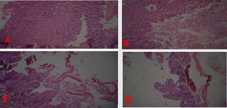
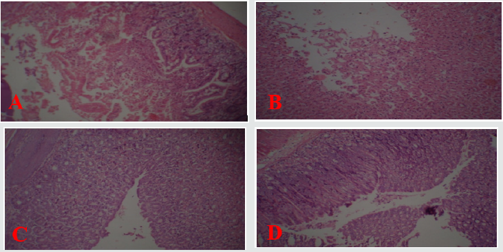
As shown in Figure 1 normal gastric mucosa Figure [A] compared with Figure [B & C] with treatment by ethanol there is exfoliation and sloughing of gastric cells with inflammatory cell infiltrate and congestion of blood vessels Figure [D]. Figure 2 shows partial improvement in gastric lesion in the group treated by oral ginger treatment at dose 300mg/kg as there is mild exfoliation of gastric cells Figure [A]. Moderate gastric protection by (I.P) treatment of ginger at a dose 600mg/kg as there is moderate exfoliation of gastric cells as shown in Figure [B]. Complete protection by chronic (I.P) treatment of ginger in a dose of 300mg/kg with no exfoliation of gastric cells as shown in Figure [C]. Rantidine has moderate protective effect as shown in Figure [D] compared with extract in Figure [C] with complete protection of gastric mucosa.

Figure 1: Light photomicrographs (10× magnifications) of hematoxylin and eosin- stained sections of stomach.

A: Normal gastric mucosa.

B, C: Ethanol treated stomach with Exfoliation and sloughing of gastric cells.

D: Ethanol treated with congestion of blood vessel.
Lupinepublishers-openaccess-complementary-alternative-medicine-journal

Figure 2: Light photomicrographs(10×magnifications) of hematoxylin and eosin-stained sections of stomach:

A: Ginger treated (IP) stomach.

B: Ginger treated (IP) stomach.

C: Chronic ginger treated (IP).

D: Ranitidine treated (IP) stomach.
Lupinepublishers-openaccess-complementary-alternative-medicine-journal

In present paper ethanol administration provoked significant gastric mucosal injuries as evidence by necrosis, exfoliation and sloughing of gastric cells, congestion of blood vessels and infiltration of inflammatory cells. Cytoprotective action of ginger has been investigated using animal models of acute gastric injury induced by necrotizing agents such as ethanol. Ethanol serves as the most common ulcerogenic agent and produced sever gastric hemorrhagic erosions Robert et al. [12]; Szabo et al. [13]. The genesis of ethanol-induced gastric lesions is multifactorial with the depletion of gastric wall mucus content as one of the involved factors. Submucosal venular constriction by ethanol and eventual injury is caused due to perturbations of superficial mucosal cells, notably the mucosal mast cells leading to release of vasoactive mediators including histamine, that cause damage to gastric mucosa Hollander et al. [14]. Accumulation of activated neutrophil in gastric mucosa may be the source of free radicals Oxygen free radical which lead to increase to lipid peroxidation and damage to cell membrane are impacted in ethanol induced gastric mucosal injury Al-Harbi et al. [15]. In addition to its direct damage of gastric mucosal cells by development of free radicals, cause solubilization of mucus constituents and depressant tissue levels of protein leading to flow stasis in gastric blood. The present paper revealed parial cytoprotection by single oral dose of ginger (300mg/kg) in mice. More effective cytoprotection was elicited by (600mg/kg I.P).

The results obtained in the present paper indicated that ginger extract possess its anti- ulcerative properties through a mechanism mainly related to acid and pepsin inhibition Chronic I.P injection of plant extract in a dose of (300mg/kg) more effective in prevention lesion formation. The mechanism of ginger extract in chronic (I.P) protection may be due to counteracting the active oxidant radicals, decreasing mucosal cell shedding and thicking the mucus membrane. The mechanism of anti-ulcer effect of ginger may be due to presence of 6-Gingersulfonic acid and three monoacyldigalactosyl gylcerols including ginger glycolipid A. B. C have been isolated from dried rhizome of Zingiber Officinale which are potent anti-ulcer components Yoshikawa et al.[16]. 6- Gingerol and 6- shogaol are two other anti- ulcer components that are less potent but are mainly responsible for ginger pungency. The results of present paper in accordance with previous reports in which water and methanolic extract of eight Zingiberaceae herbs caused a significant decrease in gastric secretion in un-anesthetized rabbits and the effect of water extract was similar to cimetidine Sakai et al. [17].


For More Lupine Publishers Open Access Journals Please visit our Website: https://lupinepublishers.us/
To Know More About Open Access Publishers Please Click on Lupine Publishers

Follow on Twitter:  https://twitter.com/lupine_online

https://lupinepublishers.com/complementary-alternative-medicine-journal/fulltext/cytoprotective-effect-of-zingiber-officinale-extract-on-alcohol-induced-gastric-lesion-in-mice.ID.000139.php

Lupine Publishers | Tongluo Decoction Blocks Apoptotic Mitochondrial Pathway and Inhibits Cardiomyocyte Apoptosis of Coronary Microembolization Rats

 Lupine Publishers| Open access Journal of Complimentary and Alternative Medicine 


BackgrBackground: We found that Tongluo Decoction has had good protection for myocardial cells injury of CME rats. But it is not clear whether Tongluo Decoction has inhibitory effect on myocardial apoptosis. So we want to study the effect and mechanism of Tongluo decoction to cardiomyocyte apoptosis following coronary microembolization in rats. Methods: 46 male Wistar rats were randomly divided into blank group (n=6), model group (n=10), Tongluo Decoction low dose group (n=10), Tongluo Decoction high dose group (n=10) and Tongxinluo group (n=10). In addition to the blank group, the rats of other groups were established the model of autologous thromboembolism microembolism by interventional method. The low dose group of Tongluo Decoction was given 7.8g/kg for gavaging, the high dose group of Tongluo Decoction was given 23.4g/kg, Tongxinluo group was given 0.2g/kg, once-a-day, for 12 day. The judgment of the model was done with HE and HBFP staining, the myocardial apoptosis rate was observed by TUNEL staining. Western Blot was used to detect Bcl-2, Bax and Caspase-3 in cardiac myocytes.

Results: The microembolization vessel count ratio, ischemic myocardial area ratio and the apoptosis rate of myocardial cells in model group were significantly higher than that in blank group, bcl-2 and bcl-2/bax decreased, and bax and caspase-3 were significantly increased (P < 0.05).Compared with model group, Tongluo Decoction low dose group, Tongluo Decoction high dose group and Tongxinluo group microembolization vessel count ratioischemic myocardial area ratio apoptosis rate decreased (P < 0.05), bcl-2, bcl-2/bax increased, Bax and caspase-3 decreased (P < 0.05). Compared with Tongxinluo group, the ratio of ischemic myocardial area, microembolization vessel count ratio, apoptosis rate, the expression of bax and caspase-3 proteins decreased (P > 0.05), bcl-2 and bcl-2/bax ratio increased in Tongluo Decoction high dose group (P > 0.05). bcl-2 and bcl-2/bax ratio increased obviously in Tongluo Decoction low dose group (P < 0.05).

Conclusion: Tongluo Decoction can inhibit cardiomyocyte apoptosis following coronary microembolization in rats, and its possible mechanism is related to blocking mitochondrial apoptotic pathway.

Keywords: Tongluodecoction; Rat; Coronary microembolization; Cardiomyocyte apoptosis

Abbrivations: Percutaneous coronary intervention (PCI); Coronary microembolization (CME); Hematoxylin-Eosin staining (HE); Hematoxylin basic fuchsin picric staining (HBFP); Terminal deoxynucleotidyl transferase-mediated dUTP Nick End-Labeling (TUNEL); High performance liquid chromatography (HPLC); Tongluo decoction (TLD); Optical density (OD); Bcl-2 Assaciated X protein (Bax); Cysteinyl aspartate specific protease-3 (Caspase-3); B-cell lymphoma-2 (Bcl-2)

Introduction

In the process of percutaneous coronary intervention which is used to treat coronary heart disease, the small fragments that come from mechanical injury of coronary atherosclerosis, with the flow of blood circulation to the distal coronary artery, block the distal microvascular, which leading to the obstruction of coronary microvascular circulation, that is coronary microembolization(CME). CME is a serious complication of PCI, which can cause myocardial ischemia and cardiac dysfunction, and even lead to death. Numerous experimental studies [1, 2] have shown that apoptosis plays an important role in CME lesions. The research showed that Chinese materia medica can improve myocardial injury caused by coronary microembolization [3]. We found that Tongluo Decoction has had good protection for myocardial cells injury of CME rats. The Tongluo decoction could inhibit the pathological change of CME in rats, reducing the ingury of myocardial cells, inhibiting the expression of inflammatory cytokines and promoting the microemboli vascular recanalization [4]. But it is not clear whether Tongluo Decoction has inhibitory effect on myocardial apoptosis of CME rats. Therefore, in this study, we established coronary artery microembolization model of rats by injecting autologous thrombus through the right common carotid artery by interventional method with balloon catheter [5]. The effect and mechanism of Tongluo Decoction on cardiomyocyte apoptosis following CME in rats were studied.

Methods

Preparation and quality control of tongluo decoction

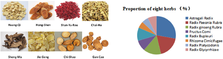
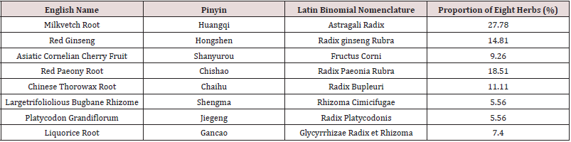
Tongluo Decoction mainly consisted of eight herbs: [6- 8] The composition and proportion of herbs are shown in the diagram (Figure 1, Table 1). The herbs was purchased from the Hui ren Tang Pharmaceutical Company, and they were identified by Jianyin Li, a teacher of Lanzhou University school of pharmacy.(The voucher specimen numbers of the stored herbarium specimen are 62032120180807063LY for Astragali Radix, 620031220010918321LY for Radix ginseng Rubra, 62062319980910435LY for Fructus Corni, 62032120180811156LY for Radix Paeonia Rubra, 62032120180807062LY for Radix Bupleuri, 62062320130815322LY for Rhizoma Cimicifugae, 62032120180805049LY for Radix Platycodonis, 62032120180812164LY for Radix Glycyrrhizae. ) This herbs that was conformed to the prescription, all of them were qualified, decocted, concentrated, and then put in refrigerator at 4. The chromatographic conditions were Diamonsil C18 (2) column (200×4.6mm, 5μm), the liquid phase of the calycosin glycoside (batch number 111920-201606, National institutes for food and drug control) mobile phase: acetonitrile -0.2% formic acid solution (15:85), flow velocity:1ml/min, column temperature: 30, detection wavelength: 260nm, sample size: 10μl. Paeoniflorin (batch number X27F8C30162, Shanghai source leaf Biological Technology Co., Ltd.) mobile phase: acetonitrile-0.1% phosphoric acid solution (16:84). Flow velocity, column temperature and sample size remain unchanged. Detection wavelength: 230nm; Ginsenoside Rb1 (batch number Z06M8L30693, Shanghai source leaf Biotechnology Co., Ltd.) mobile phase: acetonitrile -0.1% phosphoric acid solution, gradient elution. Flow velocity, column temperature and sample size remain unchanged. The detection wavelength: 203nm. To calculate the content of calycosin glycoside, paeoniflorin and ginsenoside Rb1.

Figure 1: Composition of main herbs and their proportion in Tongluo Decoction.

Lupinepublishers-openaccess-complementary-alternative-medicine-journal

Table 1: The full scientific species names of ingredients of Tongluo Decoction.

Lupinepublishers-openaccess-complementary-alternative-medicine-journal

Preparation of autologous thrombus suspension

1ml blood was collected from the caudal vein of each rat , then drying, the dry blood was grinded into powder like particles. The thrombus particles with a diameter between 41μm and 80μm were mixed into a suspension, and 2×10⁵ thrombus particles were injected into the the aortic root through the right common carotid artery for establishing coronary artery microembolization model.

Animals grouping and model establishment

46 SPF healthy adult male Wistar rats (weight 350g±30g) were randomly divided into blank group (n=6), model group (n=10), Tongluo Decoction low dose group (n=10), Tongluo Decoction high dose group (n=10), Tongxinluo group (n=10). The experimental animals were purchased from the experimental animal center of Lanzhou University. The laboratory animal license number SCXK (Gan) 2013-0002. The experimental process adhered to the ethical regulations of animal experiments. The rats were anesthetized with 0.4% pentobarbital sodium solution, separated the right common carotid artery and cut the “V” incision at its distal end. The balloon catheter, which was soaked with heparin sodium saline, was pushed to the root of the aorta (about 4cm from the incision of right common carotid artery to the aortic root ), and the blood flow of the ascending aorta was blocked for 3-5s, rapidly withdrawing the guide wire during the period, and the autologous thrombus suspension was injected. Balloon catheter was pulled out, the proximal heart segment of the right common carotid artery was ligated. Penicillin 4×10⁵ U/each was given for 3 days after the operation to prevent infection.

Drug intervention

The daily dosage of the low dose group of Tongluo Decoction was calculated according to the conversion ratio of the 1:6.25 in the rats [5]. The dosage of the high dose group of Tongluo Decoction was 3 times that of the low dose group of Tongluo Decoction. Tongxinluo capsule (Shijiazhuang Yiling pharmaceutical Limited by Share Ltd) uses 9grains/day dose, dissolve it in double steamed water and intragastric gavage. Tongxinluo capsule is purchased from Hui Ren Tang Pharmaceutical Company and its specification is 0.26g/ granules. The rats of each group were given the corresponding drug by intragastric gavage for 12 days. During the test, 3 rats died in each of the model group, low dose group of Tongluo Decoction, high dose group of Tongluo Decoction and Tongxinluo group during the modeling process, and 1 died in Tongxinluo group after gavage.

Collection of heart specimens

After the drug intervention, all rats were sacrificed by cervical dislocation under anesthesia by intraperitoneally injected 0.4% sodium pentobarbital solution 1ml per 100g body weight. Then rat heart was collected. The apex of heart was fixed in 4% polycondensation formaldehyde solution. After dehydration and paraffin embedded, 5μm consecutive paraffin sections were used to HE and other staining. The base of the heart was immediately placed in a pre-freezing storage tube, stored at liquid nitrogen, and used to detect cardiomyocyte apoptosis protein by Western Blot.

HE and HBFP staining of rat myocardium

After the apex staining of heart paraffin section of rats in each group with HE and HBFP, the staining sections were observed by three researchers under 400× and 100× microscope respectively to calculate the microembolization vessel count ratio, ischemic myocardial area ratio.

Detection of cardiomyocyte apoptosis by TUNEL

The pathological tissue section of the rat in each group was treated with anti-shedding. According to the TUNEL instructions (Boster Co., Ltd., Wuhan) to stain. The number of cardiomyocyte apoptosis and the number of total cells under the microscope were observed by three researchers under 400× microscope, and the apoptosis rate of myocardial cells was calculated and the apoptosis level of cardiac myocytes was evaluated. Apoptosis rate (%)=(the number of apoptotic cells/the total cell number under microscope)×100.

Western Blot was used to detect the expression of cardiomyocyte apopt-osis proteins

To extract the protein of myocardial tissue, according to the instructions of the kit (Solarbio Science and Technology Co., Ltd. Beijing). The OD value was measured, and the standard protein curve was drawn. Then calculating the protein concentration. Prepare 12% separation glue and 5% concentration glue, 160 V constant pressure electrophoresis 100min, and PVDF membrane was then transferred, followed by incubation for 2h with blocking buffer containing 5% Non-fat milk powder. Diluting and putting relevant primary antibodies, bcl2, bax, caspase3, and GAPDH respectively (Biosynthesis biotechnology Co., Ltd. Beijing), with gentle shaking overnight at 4. Afterwards, the membrane was incubated with secondary antibody(purchased from SAB) for 1h at room temperature, then washing and exposing.

Data statistics method

The SPSS 22.0 statistics software was used to analyze the data, and the measurement data were expressed with mean±standard deviation (x±s). Single factor variance( one-way ANOVA) was used in comparison of multiple groups. And P < 0.05 indicated that the difference was statistically significant.

Results

Quality control results of tongluo decoction

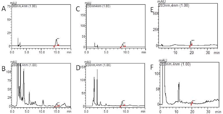
As shown in Figures 2(a)-2(f), the separation of the components to be tested is good and has no interference with other components. High performance liquid chromatography (HPLC) shows the peak appearance time of the calycosin glycoside standard and Tongluo Decoction for test sample was 15.0±0.5 min, while the paeoniflorin standard and Tongluo Decoction for test sample was 9.0±0.5 min, and the ginsenoside Rb1 standard and Tongluo Decoction was 20.0±0.5 min, and the effective components of three herbs are compared with their respective standard products, some impurity peaks appeared. However, the separation efficiency of the effective components of the three herbs were not affected. The above results indicated that the effective components of Tongluo Decoction contained of calycosin glycoside, paeoniflorin and ginsenoside Rb1. The results showed that the effective components of Tongluo Decoction included calycosin-7-glucoside, paeoniflorin and ginsenoside Rb1, and the quality of the decoction was stable and could be used for the follow-up drug intervention test.

Figure 2:

a) High performance liquid chromatography of Tongluo Decoction, calycosin-7-glucoside standard.

b) High performance liquid chromatography of Tongluo Decoction. A calycosin-7-glucoside standard, Tongluo Decoction, calycosin-7-glucoside.

c) High performance liquid chromatography of Tongluo Decoction,paeoniflorin standard.

d) High performance liquid chromatography of Tongluo Decoction, Tongluo Decoction, 1 is paeoniflorin.

e) High performance liquid chromatography of Tongluo Decoction, ginsenoside Rb1 standard.

f) High performance liquid chromatography of Tongluo Decoction, Tongluo Decoction, 1 is ginsenoside Rb1.

Lupinepublishers-openaccess-complementary-alternative-medicine-journal

HE staining results of rat myocardium

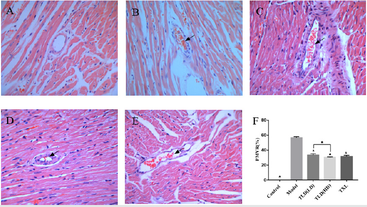
As shown in Figures 3(a)-3(f), the microembolus were not found in the microvessels of the coronary artery in the blank group. A large number of microembolus were found in the coronary microvessels in the model group, and around the microembolus there were inflammatory cell aggregation. Some of microembolus were found on the low dose group of Tongluo Decoction, the high dose group of Tongluo Decoction, and the Tongxinluo group in the coronary microvessel. The positive microembolization vessels count ratio (PMVR) in the model group was significantly higher than that in the control group (P < 0.05). Compared with the model group, the low dose group of Tongluo Decoction, the high dose group of Tongluo Decoction and the Tongxinluo group were significantly lower than those in the model group (P < 0.05). Compared with the Tongxinluo group, the PMVR in the Tongluo Decoction low dose group was increased while the Tongluo Decoction high dose group was decreased(P > 0.05). The PMVR of high dose group of Tongluo Decoction was lower than that in the low dose group of Tongluo Decoction (P < 0.05). (Figures 4(a)-4(f)).

Figure 3: Comparison of HE staining of myocardial positive microembolic blood vessels of rats in each group(400×). A blank group, B model group, C TLD(LD) group, D TLD(HD) group, E TXL group. F Comparison of HE staining of myocardial positive microembolic blood vessels of rats in each group. (▲)P<0.05, versus model group; (★)P<0.05, TLD(LD) group versus TLD(HD) group. PMVR:Positive microembolization vessel count ratio.

Lupinepublishers-openaccess-complementary-alternative-medicine-journal

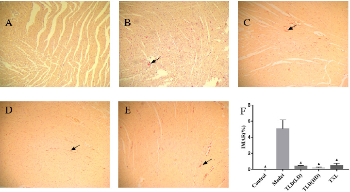
Figure 4: Comparison of HBFP staining of ischemic myocardium of rats in each group(100×). A blank group, B model group, C TLD(LD) group, D TLD(HD) group, E TXL group, F Comparison of HBFP staining of ischemic myocardium of rats in each group. (▲)P<0.05, versus model group. IMAR:Ischemic myocardial area ratio.

Lupinepublishers-openaccess-complementary-alternative-medicine-journal

HBFP staining results of ischemic myocardium

In Figures 5(a)-5(f), almost no red myocardium tissues was found in the blank group. A large number of red myocardium tissues were found in the model group. Tongluo Decoction low dose group, Tongluo Decoction high dose group and Tongxinluo group found some red myocardium tissues. Compared with the blank group, the ratio of ischemic myocardial area ratio in the model group was significantly higher (P < 0.05). Compared with the model group, the ratio of ischemic myocardial area ratio in Tongluo Decoction low dose group, Tongluo Decoction high dose group and Tongxinluo group decreased significantly (P < 0.05). Compared with Tongxinluo group, the ratio of ischemic myocardial area ratio(IMAR) in Tongluo Decoction low dose group, Tongluo Decoction high dose group was decreased, but the difference was not statistically significant (P > 0.05) .

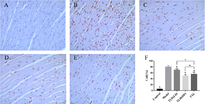
Figure 5: Comparison of TUNEL staining(400×)and apoptosis rate of myocardial cells in each group of rats. A blank group, B model group, C TLD(LD) group, D TLD(HD) group, E TXL group. F Apoptosis rates of cardiomyocytes in TUNEL staining groups were compared, (▲)P<0.05, versus model group; (#)P<0.05, TLD(LD) group versus TXL group; (★)P>0.05, TLD(HD) group versus TXL group. CAR: cardiomyocyte apoptosis ratio.

Lupinepublishers-openaccess-complementary-alternative-medicine-journal

Detection of cardiomyocyte apoptosis by TUNEL

As shown in Figure 6, TUNEL staining showed that the normal nuclei were pale blue, the apoptotic nuclei were tan, nuclear chromatin was concentrated, and the nuclei were broken into unequal nucleus fragments, accompanied by the formation of apoptotic bodies. There were almost no tan nuclei in the control group, and a number of tan nuclei in the model group. Tongluo Decoction low dose group, Tongluo Decoction high dose group and Tongxinluo group have some tan nuclei. The apoptosis rate of myocardial cells in the model group was significantly higher than that in the control group (P < 0.05). Compared with the model group, the cardiomyocyte apoptosis rate in Tongluo Decoction low dose group, Tongluo Decoction high dose group and Tongxinluo group decreased significantly (P < 0.05). Compared with Tongxinluo group, the decrease of cardiomyocyte apoptosis rate in Tongluo Decoction low dose group was small (P < 0.05) , but the decrease of cardiomyocyte apoptosis rate in Tongluo Decoction high dose group was much more (P > 0.05).

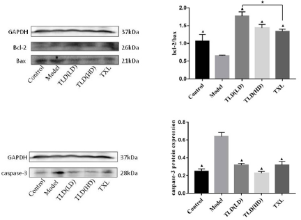
Figure 6: Western Blot detection of bcl-2、bax、caspase-3 protein expression in cardiac myocytes.

Lupinepublishers-openaccess-complementary-alternative-medicine-journal

Western Blot detect the expression of Bcl-2, Bax and caspase-3

Protein in Cardiac Myocytes

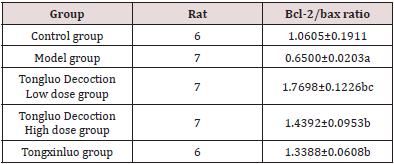
As shown in Figures 3(a)-3(f)) and (Table 2), compared with the control group, the expression of Bcl-2 protein and bcl-2/bax of the model group were decreased, the expression of Bax and Caspase-3 in the model group were significantly increased (P < 0.05). Compared with the model group, the expression of Bcl-2 protein, bcl-2/bax were increased and the expression of Bax and Caspase-3 were decreased in the Tongluo Decoction low dose group, Tongluo Decoction high dose group and Tongxinluo group (P < 0.05). Compared with Tongxinluo group, the expression of Bcl-2 protein, bcl-2/bax increased (P < 0.05), the expression of Bax and caspase-3 protein decreased in the Tongluo Decoction low dose group and the Tongluo Decoction high dose group (P > 0.05). These results indicate that the coronary microembolization(CME) can induce cardiomyocyte apoptosis, and Tongluo Decoction and Tongxinluo can inhibit cardiomyocyte apoptosis.

Table 2: The results of bcl-2/bax ratio in each group of rats (x ± s ).

Lupinepublishers-openaccess-complementary-alternative-medicine-journal

Note: Comparison with control group.

aP﹤0.05; Comparing with model group.

bP﹤0.05; Comparing with Tongxinluo group, cP﹤0.05.

Discussion

CME is an common complication in the interventional therapy of coronary heart disease. It is one of the important reasons for the occurrence of “no reflow” and “slow flow” of coronary artery. It is an important factor of poor prognosis and even death in some patients after PCI. The secondary myocardial apoptosis and necrotic myocardium irreversible injury are independent predictors of no reflow and poor prognosis. Research shows [4] that a stable, reliable and economical model of coronary arteria microembolism can be established by injecting autologous thrombus to the aortic root of rat through the right common carotid artery. In this study, we followed this method and established the rat CME model successfully. After the drug intervention 12 days, we found that both Tongluo Decoction and Tongxinluo could contribute to the narrowing of the microembolus, promote the blood circulation of the coronary microembolic vessels and improve the condition of myocardial ischemia. Tongluo Decoction is composed of many kinds of Chinese medicinal materials according to the rule of monarch(Jun), minister(Chen), assistant(Zuo) and guide(Shi). It has the effects of benefiting qi for activating blood circulation and disperse blood stasis and dredge collateral circulation. Among this Chinese medicine compound, Astragali Radix(Huangqi) as the monarch, compatible with Radix ginseng Rubra (Hongshen) can tonify Qi of spleen and Lung significantly. It made QI and blood own a recourse to metaplasia. Compatibility of Radix Paeonia Rubra (Chishao) have an effect on blood stasis runs and relieve pain, cooling blood and subsidence of a swelling. Assistance of Radix Bupleuri(Chaihu), Rhizoma Cimicifugae(Shengma), Radix Platycodonis(Jiegeng) for lifting Yangqi and carrying medicine upgoing. Qi running leads to blood circulation. Then add Fructus Corni(Shanyurou) to nourish the Yin of liver and kidney. Radix Glycyrrhizae(Gancao) as guide plays an harmonization of all drugs role in the compound. All drug compatibility, reflect a efficacy of tonifying qi and yin and promoting blood circulation to remove meridian obstruction. Zhang Zhenguo and other studies [6] found that Tongluo Decoction could inhibit the pathological process of coronary microembolism, reduce myocardial damage, inhibit the expression of inflammatory factors and promote the role of microembolic vascular recanalization.

Apoptosis is an active and orderly death process, which is produced by the body in the physiological and pathological conditions to maintain the homeostasis of its own internal environment [7]. Studies have shown that apoptosis is one of the possible mechanisms of ventricular dysfunction induced by coronary microembolization [8]. Anti apoptotic therapy can significantly inhibit the apoptosis of cardiomyocytes after CME in rats [9-11]. Studys showed that Tongxinluo capsule could inhibit cardiomyocyte apoptosis [12, 13]. In our study, Tongxinluo was used as a positive control group to further study the mechanism of Tongluo Decoction from the perspective of apoptosis. Mitochondria mediated apoptosis pathway is one of the most important apoptotic pathways. Numerous studies have shown [14-16] that inhibition of mitochondrial mediated apoptosis plays an important role in improving CME induced myocardial damage and cardiac dysfunction. The regulation of mitochondrial apoptosis is mainly mediated by the regulation of Bcl-2 family proteins [17, 18]. After the destruction of mitochondria, the release of cytochrome C increased. Under the regulation of Bcl-2 family protein, the release of cytochrome C was induced by anti apoptotic members and inducing apoptotic members, eventually causing the activation of Caspase-3, resulting in the apoptosis [19]. Cysteinyl aspartate specific protease (caspase) is the main effector factor of apoptosis.

Wang JiangYou and other studies [20] showed that increased the expression of Caspase-3 in rat cardiomyocytes after CME, and pretreatment with atorvastatin could downregulate the expression of Caspase-3. Liu Tao and other studies [21] found that after CME bcl-2 expression decreased, Bax and Caspase-3 expression increased, silence LOX-1 can reverse Bcl-2, Bax, caspase-3 protein expression. Bcl-2 can inhibit apoptosis, while Bax can promote apoptosis [22]. The ratio of Bcl-2/Bax is an important parameter of mitochondrial signaling pathway, and the ratio between them is one of the important causes of apoptosis [23, 24] Our study showed that the apoptosis rate of myocardial cells in the CME model group was significantly increased. The expression of Bcl-2 protein, bcl-2/bax were decreased and the expression of Bax and Caspase-3 were significantly increased in the model group. It was indicated that CME induced the cardiomyocytes apoptosis. The apoptosis rate of cardiac myocytes in Tongluo Decoction low dose group, Tongluo Decoction high dose group and Tongxinluo group was significantly lower than that of model group (P < 0.05), and the expression of Bcl-2 protein and bcl-2/bax increased, and the expression of Bax and caspase-3 protein decreased (P < 0.05). It indicates that Tongluo Decoction and Tongxinluo capsule can inhibit the cardiomyocytes apoptosis of rat after CME. Compared with Tongxinluo group, the degree of decrease on cardiomyocyte apoptosis rate in Tongluo Decoction low dose group was lower (P < 0.05) while Tongluo Decoction high dose group was higher (P > 0.05). The results of Western Blot showed that, compared with Tongxinluo group, the expression of Bcl-2 protein, bcl-2/bax increased (P < 0.05), the expression of Bax and caspase-3 protein decreased in the Tongluo Decoction low dose group and the Tongluo Decoction high dose group (P > 0.05). It shows that Tongluo Decoction is almost equivalent to Tongxinluo capsule in the treatment of CME, which can inhibit the apoptosis of myocardial cells of rats after CME, and its mechanism may be related to the blocking of mitochondrial apoptosis pathway.

Conclusion

Tongluo Decoction may inhibit the cardiomyocytes apoptosis of CME rats by blocking mitochondrial apoptosis pathway and protect their myocardium.

For More Lupine Publishers Open Access Journals Please visit our Website:
To Know More About Open Access Publishers Please Click on Lupine Publishers

Follow on Twitter:  https://twitter.com/lupine_online

https://lupinepublishers.com/complementary-alternative-medicine-journal/fulltext/tongluo-decoction-blocks-apoptotic-mitochondrial-pathway-and-inhibits-cardiomyocyte.ID.000138.php

Tuesday, September 8, 2020

Lupine Publishers | The Effect of Quran Vocalizations on Pain Relief in Neonates During Treatment Procedures

Lupine Publishers | Open access Journal of Complimentary and Alternative Medicine 


Pain is one of the most common human problems that has been experienced more or less in all people but the symptoms of it are usually different in different ages [1]. One of the most common misconceptions is that infants do not feel pain because their nervous system has not developed enough. No longer is the experience of pain painful and harmful for infants because they lack memories of pain [2]. Recent research has provided ample evidence that infants, even preterm infants, have a more mature central nervous system than might be thought. The onset of neural pathways myelinated in the fetus is during the second and third trimesters of the fetus, but may begin earlier. Fine or non-myelinated fibers also transmit the stimulation of pain, but only slower the rate of transmission [3]. Stress from pain, even brief and brief, can lead to problems such as (hypermetabolic status, cardiac arrhythmias) and even complications and delay in neonatal recovery. In infants, behavioral and physiological symptoms of pain are the only way to express it. For this reason, less research has been done to try and quantify neonatal pain and to facilitate the identification of pain intensity by nurses [2]. In study by Poznanski, infants responded to circumcision ISSN: 2644-1217 DOI: 10.32474/OAJCAM.2020.02.000137 Abstract Introduction: Pain is a destructive one and remains in the mind of the child suffering from it Pain management in newborns through pharmaceutical and complementary medicine, including Quran recitation is done. Hence the aim of this study was to effect of Quran vocalizations on pain relief in neonates during treatment procedures in Imam Hossain hospital of Shahroud in 2014. Methods and Materials: This cross-sectional study that compared with study of Quran audio impact in reducing neonatal pain during medical procedures performed. Sample size was estimated to be about 120, and this number of the subjects was selected in six months period. The data were recorded in check list prepared according to also, theory of development and Lawrence s NeonateInfant pain scale [NIPS]. Results: In the intervention group, 28 patients (46.7%) were female and the rest were male while 29 patients in the control group (48.3%) were male and the rest female that were not significant differences. The results of this study showed that the experimental group, the mean number of breathing, crying, movements of arms and legs as well as the mean state of consciousness at the end of treatment compared to baseline that this differences were significant (p<0.001), while in the control group this difference was not seen. So the results showed that the highest impact Quran was the end of the procedure started 10 minutes to reach its maximum impact. Conclusion: According to the results can be obtained from more appropriate to examine the behavioral pain symptoms of physiological performance of this tool to promote and Quran recitation to reduce pain in newborns help, but it is necessary to determine the duration of the sound of heavenly research did more. Keywords: Pain; Neonatal; Quran vocalizations; Behavioral sign’s; Treatment procedure Citation: Pouneh Z, Maryam F, Elahe E, Elahe Y, Mohammad B S. The Effect of Quran Vocalizations on Pain Relief in Neonates During Treatment Procedures. Open Acc J Comp & Alt Med 2(3) - 2020. OAJCAM.MS.ID.000137. DOI: 10.32474/OAJCAM.2020.02.000137. Open Acc J Comp & Alt Med Volume 2 - Issue 3 Copyrights @ Mohammad Bagher Sohrabi, et al. 153 pain with screams and limb movements [4]. Other studies have also shown that in neonates, pain reactions can occur in the form of leg pulling, upper and lower limb flexion, facial tearing, and crying [2, 4]. Also in Kuner & Fuller study, it was found that nurses’ work experience and experience are directly related to their diagnosis and rating of neonatal pain [5]. Nurses’ experience and skills, especially those working in the neonatal ward, are vital. It was also found that these pain symptoms in infants can be altered by altering the vital signs of their infants, so that Beaver has shown in a study that elevated heart rate and blood pressure and is decreasing in blood oxygen levels indicate pre-term neonatal pain [6]. Whichever of these diagnostic symptoms is present, neonatal pain should be controlled as soon as possible after diagnosis. Different methods such as drug therapy, massage therapy and complementary medicine are used to control neonatal pain. One of the most important methods of complementary medicine is music and vowel therapy and one of the most beautiful sounds is the beautiful sound of reciting the verses of the Holy Quran with its own order which is considered as one of the most magnificent aspects of the Holy Quran miracle [7]. In the verses of the Holy Quran, the letters are uniquely aligned, so that when they hear, there is no rhythm or weight, no rhythm or weight, no rhythmic rhythm is heard, so that no text is loud Reading, like the Holy Quran, is not compact and effective [8]. In Iran, many studies have been done on the effect of the Quran’s sound on patients in various fields and positive results have been obtained. For example, the results of the study [7], to investigate the effect of the Holy Quran on vital signs of patients before cardiac surgery showed a significant difference in heart rate and respiratory rate in the intervention group compared to the control group [9]. In Khatoni study to investigate the effect of the holy Quran on the level of anxiety in patients admitted to ICU in one of Tehran hospitals, Khatoni concluded that listening to the Holy Quran may reduce anxiety in patients [10]. Regarding the necessity of neonatal pain control when performing medical and therapeutic procedures to prevent neurobehavioral complications and given the positive effects of Quran phonics on the control of physical and psychological problems in a number of patients and also because there has been no study on the effect of the Holy Quran on pain control during neonatal treatment procedures, the aim of this study was to effect of Quran vocalizations on pain relief in neonates during treatment procedures in Imam Hossain hospital of Shahroud in 2014. Materials and Methods This study was a case-control study that was conducted in the first half of 2014. The study population included 120 neonates admitted to the neonatal ward of Imam Hossein hospital in Shahroud. In this study continuous sampling method was used. The researcher, after obtaining the necessary authorization for the research from Shahroud University of Medical Sciences, was present in the study environment for several consecutive days and selected samples from the neonates who were eligible to participate in the study. A written letter from parents was included in the study. Samples were randomly divided into two groups using four randomized blocks. The children were divided into two equal case groups (Quran voice broadcasting) and control groups (without Quran voice broadcasting). Inclusion criteria for neonates included: Iranian nationality, fetal age 34-38 weeks at birth, weight over 2500 g, Apgar score of 7 and above at first and fifth minutes of birth, lying in bed, breast feeding, evidence of auditory system health and no history of severe response to sound stimulation in nursing reports, lack of intrauterine growth retardation, absence of anomalies and nervous system problems (hydrocephalus, microcephaly, meningo mycelium, intrahepatic rheumatoid arthritis, spina bifida, cerebral palsy), absence of acute disease (neonatal infection, asphyxia, severe hypoxia, intestinal necrosis, acute pulmonary disease), anemia and lack of sedative drugs such as phenobarbital and my mother had a history of addiction to drugs. Exclusion criteria included: Severe reaction to voice stimulation during intervention [including: hiccup symptoms, tilted mouth, eye squeeze, eye rolling, tongue twitching, finger opening, motor scrambling, crying, screaming and stuttering], abnormal physiological parameters such as heart rate greater than 200, reduction in arterial oxygen saturation were less than 80% during the intervention, and infant’s need for any medical or nursing or touch intervention during the study. For the infants of the case group while performing the medical procedures, the Quran voice transmission via A4TECH model EST2010 was used. The sound level of the audio player was determined in dBel by the sound level meter and under expert supervision. For the control group no Quranic sounds were broadcast. Before the intervention, the infant’s medical record was reviewed by the researcher and demographic information including variables such as type of delivery, date of birth, sex, gestational age, chronological age (number of days past the baby’s birth), birth weight, inclusion weight and minute Apgar The first and fifth were recorded in the data collection form. Then the neonate’s hearing was confirmed by observing the startle reflex and response to the acoustic stimulus and the baby’s weight was determined by digital scales. Before attaching the baby to the monitor, the researcher made sure that the baby received, changed, and needed no nutritional intervention and care during the next 40 minutes. Then, the hands were washed, the infant was laid back on the couch, and hair neutrodes were attached to the infant for evaluation of physiological indices. The oxygen saturation electrodes were mounted on the anterior and posterior surfaces of the foot and covered with carbon to increase the accuracy of the device and to prevent light effects. To record heart rate and respiratory rate, three electrodes were mounted on the infant’s chest in a triangular fashion and fixed with anti-allergic adhesive to prevent displacement during physiological index recording. To reduce infant manipulation, the indices were measured continuously and only the information recorded at the desired time was used to calculate and compare differences between groups. Earphones were used to prevent the effects of sound on other neonates during audio playback. The handset was connected to the MP3 Player, the volume was controlled on the device, and then the handset was placed on the baby’s ear. After the baby was no longer Open Acc J Comp & Alt Med Volume 2 - Issue 3 Copyrights @ Mohammad Bagher Sohrabi, et al. Citation: Pouneh Z, Maryam F, Elahe E, Elahe Y, Mohammad B S. The Effect of Quran Vocalizations on Pain Relief in Neonates During Treatment Procedures. Open Acc J Comp & Alt Med 2(3) - 2020. OAJCAM.MS.ID.000137. DOI: 10.32474/OAJCAM.2020.02.000137. 154 in need of touch or manipulation, information recording began. Physiological responses were recorded every 10 minutes in three stages immediately before the intervention, during the intervention (10-20 minutes after the intervention) and 10 minutes after the intervention. The first stage of recording the infant’s physiological responses was performed just before the infant started and did not manipulate based on the numbers that the monitor displayed. Then, if the infant belonged to the intervention group, the Holy Qur’an’s voice would begin to be broadcast to him and he would continue to record physiological responses at 10-minute intervals. The Quran sound was transmitted to the intervention group for 20 minutes and at 20 minutes after recording the physiological responses, the sound player was switched off and the last recording was performed 10 minutes after the mute. The infants in the control group, like the intervention group, were attached to the monitor and their physiological responses were recorded every 10 minutes. In order to eliminate the confounding effect of headphones in the control group, infants were put on earphones, with the exception that no sound was delivered. Room temperature was maintained in the range of 27-28 °C. All infants in both groups were followed for 6 months during medical procedures including blood sampling, venipuncture, intramuscular injection, gastric catheterization, and cerebrospinal fluid (LP) ingestion. Instruments for measuring changes included neonatal balance, cardiac monitor, arterial oxygen saturation device, and continuous monitoring of experienced nurses during procedures. Simultaneous observation was used to avoid measurement error. Two trained colleagues were asked to simultaneously monitor the heart rate, respiratory rate, and oxygen saturation percentage at the desired time on the monitor and to record in separate questionnaires for each infant. The mean of these numbers was then used to report changes in each infant. Neonatal pain assessment tools have been used to assess behavioral symptoms of neonatal pain. This tool (Neonatal Infant Pain Scale) consists of 6 options that only have zero, one, and two scoring options, and five options, namely face states, respiratory patterns, hand movements, leg movements and level of consciousness, with scores of zero and one. The score range will be between zero and seven. These changes are recorded in the specific form of each baby. After collecting, the isolates were entered into SPSS 16 software and analyzed by statistical tests. In case of nonnormality of variables, Mann-Whitney test was used to compare the means between the two groups and Wilcoxon was used to compare means within groups. In normal distribution, independent t-test was used to compare the means between the two groups. Parents of infants who participated in the study were provided with complete information about the research and its aims and importance. Parents were assured that participation in the study was voluntary and not participating in the study would not affect their infant care services and information, and that information would be kept confidential. If parents wish to participate in the study, a consent form is provided for them to read and sign. Results Of the 120 neonates who were randomly divided into two equal groups of case and control, the demographic and contextual information (gender, type of delivery, neonatal apgar score at 1 and 5 minutes at birth, fetal age, age, birth weight and neonatal weight at the time of study) no significant difference was observed between two groups. These results are shown in Table 1. Based on independent t-test in the intervention group, mean difference in breathing rate, crying intensity, hand and foot movements and also in Mann-Whitney test mean difference in consciousness status at the end of the intervention was significantly different from the baseline level (p <0.001). This difference was not observed in the control group. The results are shown in Table 2. So the results also showed that the maximum effect of the Quranic phoneme started at 10 min and reached its maximum effect at the end of the procedure (min of 20). Table 1: Demographic characteristics of neonates in two groups at baseline. Variable Intervention group (n=60) Control group Gender (girls / boys) 32/28 31/29 0.213 Type of Delivery (Normal / Cesarean) 41/19 39/21 0.178 Apgar minute 1 8.17±0.45 8.23±0.59 0.235 Apgar minute 5 9.41±0.75 9.35±0.68 0.088 Fetal Age (Week) 33.28±5.12 34.08±4.88 0.109 Calendar Age (days) 3.85±4.25 4.10±4.30 0.075 Birth Weight (g) 2750±350 2690±438 0.146 Study Weight (g) 2990±523 2630±450 0.144 Table 2: Comparison of mean score of physiological variables and mean state of consciousness at the end and baseline in the intervention and control groups. Points Intervention group (n=60) Control group Before intervention 6.4±1.5 6.2±1.8 0.452 After intervention 3.5±3.8 5.7±0.5 0.001 Discussion The results of this study showed that there was a significant difference between the two groups in terms of changes in physiological symptoms before and after the intervention. Among the physiological symptoms, the most common symptoms were facial flares. Seymour et al., in their study suggested that nurses were more concerned with sweating and discoloration than physiological symptoms among infants [11]. The most commonly observed behavioral signs are facial changes and then changes in respiratory pattern and crying. This indicates that during behavioral procedures, changing facial expressions, crying, and changing the respiratory pattern are key clues to assess neonatal pain. Many studies have been done to evaluate the pain of infants and also how nurses and parents have used and interpreted the pain, which have yielded similar results. One of these studies is Bore et al., Which has been conducted to investigate neonatal reactions to painful stimuli. Results showed that preterm infants use more physiological symptoms, especially increasing of heart Citation: Pouneh Z, Maryam F, Elahe E, Elahe Y, Mohammad B S. The Effect of Quran Vocalizations on Pain Relief in Neonates During Treatment Procedures. Open Acc J Comp & Alt Med 2(3) - 2020. OAJCAM.MS.ID.000137. DOI: 10.32474/OAJCAM.2020.02.000137. Open Acc J Comp & Alt Med Volume 2 - Issue 3 Copyrights @ Mohammad Bagher Sohrabi, et al. 155 rate, respiratory rate and blood resure, and term infants use behavioral symptoms such as crying and facial changes to express pain [6]. In Jones’ study, restlessness, crying, and frustration were reported as the most behavioral symptoms of pain [12]. In their study, Throbbed and Howard stated that nurses often identified restlessness, frowning, crying, hypertension, and respiratory distress as both behavioral symptoms in term and preterm infants [2]. In both of these studies, a pain assessment tool was used, and it is noteworthy that although in the Howard and Thurber study, the neonatal intensive care unit nurses used more equipments than the Jones research, similar symptoms were observed. And the other point is that out of the selected symptoms, only the heart rate monitor needs a special device. These results suggest that nurses with the least equipment needed can use their observational skills to assess neonatal pain. Lawrence states that the standard form of infant pain assessment has high internal validity and can be used to demonstrate the severity of infant pain by referring to behavioral symptoms during the procedure and even after the procedure. The results of the present study also showed a decrease in physiological stress in the intervention group compared to the control group. While Coleman and Pratt found in a study that playing music in premature infants increased the rate of breathing and decreased heart rate [12]. A study by Calabro et al., showed that music had no significant effect on neonatal respiratory rate and heart rate [13]. According to the study by Arnon et al., The mean heart rate in the live music group was significantly lower than the two recorded and control groups. They concluded that playing live music had a greater impact on the physiological and behavioral responses of preterm infants than recorded music [14]. In a study conducted by Majidi to investigate the effect of the holy Quran on the level of stress in patients, the results showed a decrease in breathing and pulse rate after the intervention in the holy Quran compared to the control group [15]. The present study is consistent. The findings of this study are in agreement with the study of Collins, Kak and Chaw. In the study of Chaw et al., Neonates who received music therapy during endotracheal suctioning had higher oxygen saturation levels than the control group [16]. In the Cassidy and Stendley study, in neonates receiving lullaby music, the arterial oxygen saturation level was significantly higher than the control group [17-20], however, Calabrera et al., did not report a significant difference in the effect of music on the level of saturated oxygen saturation [13], which is different from the results of the present study. Also in a study by Neil et al., the effect of music and sound recorded in neonatal intensive care units was compared, with no difference in arterial oxygen saturation [2]. The differences in the results of studies that examined the effect of music on preterm infants may be due to differences in the number of samples, the use of different types of music in different ways, the effects of confounding variables, different methods of data collection, and so on. So far, no study has been done on the effect of the holy Quran on infants, but the results of this study are in line with the studies on the effect of the holy Quran on the level of anxiety and physiological responses of the patients. For example, the results of a study by Nasrabadi showed that listening to the Qur’an reduces the severity of pain and its associated behavioral symptoms in patients after abdominal surgery [17]. In another study by Majidi to investigate the effect of Quran phonation on the level of anxiety in patients admitted for angiography, the results showed a decrease in personality and situational anxiety, as well as a decrease in systolic and diastolic blood pressure, and pulse and respiratory rate in the Quran group [7]. In a study by Elder Abadi et al., to investigate the effect of the holy Quran on vital signs of patients before cardiac surgery, there was a significant difference in heart rate and respiratory rate between the intervention and control groups. They concluded that listening to the Holy Quran recalls stress- dependent physiological responses in patients [9]. Despite the contradictory findings regarding the effect of music on the physiological responses of preterm infants, the results of all studies in Iran show the positive effect of the holy Quran on reducing pain and anxiety in adults. Since most of these studies used the verses of surah Yusuf, so according to the expert advisor, verses 7-23 of surah Yusuf were chosen for this study by the voice of Master Mohammad Shayta Anwar. Undoubtedly, the use of other verses of the holy Qur’an can pave the way for further research into the effect of this heavenly calling. Music and tunes reduce the activity of the neuroendocrine and sympathetic systems and allow the parasympathetic to overcome the sympathetic, resulting in relaxation and sleep, decreased heart rate, deep breathing regulation, relaxation of the muscles and induction. The state of consciousness is calming. The neuroendocrine system is affected by music in three ways: 1-The secretion of endorphins from the pituitary gland which relieves pain and affects mood and memory. 2- Decreased secretion of catecholamines, such as epinephrine and epinephrine light from the adrenal gland, which decrease heart rate, metabolism, blood pressure, free fatty acids and oxygen consumption. 3- Decrease in adrenal corticosteroids that are released during stress [17]. Another mechanism for how music works is that music may distract the infant from distracting noise and thereby reduce the baby’s stress responses. This response has been suggested as a tool to relax the central nervous system and reduce stress responses [7]. Finally, it can be concluded that the sound of the Holy Quran may have reduced stress in neonates with the mechanism of decreased sympathetic activity and secretion of catecholamines and increased parasympathetic activity and endorphins secretion. Conclusion According to the results of this study, behavioral and physiologic observable symptoms can be used to assess neonatal pain. Of course, health care providers and nurses may also pay less attention to physiological symptoms than behavioral symptoms. But as the makers and users of pain assessment tools state, the combinations of a behavioral pain assessment form with physiological symptoms of pain increases its content validity. Since there was no negative effect on infants’ physiological parameters in the intervention group, so further research can be done to reduce stress and improve physiological status of neonates in hospitalized infants was used Open Acc J Comp & Alt Med Volume 2 - Issue 3 Copyrights @ Mohammad Bagher Sohrabi, et al. Citation: Pouneh Z, Maryam F, Elahe E, Elahe Y, Mohammad B S. The Effect of Quran Vocalizations on Pain Relief in Neonates During Treatment Procedures. Open Acc J Comp & Alt Med 2(3) - 2020. OAJCAM.MS.ID.000137. DOI: 10.32474/OAJCAM.2020.02.000137. 156 in the hospital. Limitations of this study include the inability to measure the intensity of ambient noises, the lack of a monitor with a printer, and the presence of unwanted ambient noises. 


For More Lupine Publishers Open Access Journals Please visit our Website:

https://lupinepublishers.us/

For More Complementary and Alternative medicine Articles Please Click Here: https://lupinepublishers.com/complementary-alternative-medicine-journal/index.php

To Know More About Open Access Publishers Please Click on Lupine Publishers

 Follow on LinkedIn: https://www.linkedin.com/company/lupinepublishers

 Follow on Twitter:  https://twitter.com/lupine_online

Lupine publishers| Scilit


Monday, September 7, 2020

Lupine Publishers | Failure and Stupidity

Lupine Publishers | Open access Journal of Complimentary and Alternative Medicine


If we have so consistently failed in our efforts to establish a world of peace and plenty for all, it may be because we are not trying to do that. Most people and corporations are basically out for themselves and not much interested in improving the system. Although they may be un- consciously involved in cooperative, synergistic movements to construct more complex societies, this is, at best, half of the story. For ages, people have flattered themselves with the pleasing notion that we are intelligent, God’s favorites, free, etc. Recently, analysts have carried on this tradition by emphasizing the anabolic aspects of civilization, and it is true, we can and do cooperate, and the whole can be greater than the sum of the sacrificing parts [1]. However, there is another side to the story, and it is not as flattering as that which emphasizes our constructive nature. It is a legacy of the cynics and their intellectual descendants who viewed humanity as mean, depraved, evil and stupid. Not surprisingly, scientists have been reluctant to carry on this tradition, and those who have not usually been well received. The shock and dismay that greeted Freud’s revelations are representative of the reactions of both the public and professionals to theories about human behavior that are both sound and unsettling. He has been denounced for fabricating evidence, falsifying cures and being generally wrong (especially about women), but he gave meaning to individual lives in a culture of alienation created by Nietzsche [2]. As we all know, the sad fact of life is that there is a catabolic side to nature. Civilizations both rise and fall. The same schemas which promote social cohesion can and it seems invariably do corrupt learning and adaptation and thus lead to their own disintegration. The whole becomes less than the sum of its parts as it dominates to the point of preventing subgroups from carrying out their functions effectively. Although Americans do not brag about it, the United States was peopled by failures. Their ancestors emigrated because they were or anticipated being failures in the old country. Upon arrival, they failed in farming, mining, business and battles. Crackpots invented ships that would sink, shovels that would not dig and boilers that would explode [3]. Builders constructed firetraps that were safe at any height–until they collapsed. As development progressed, slums arose in the cities while in the country, land was cleared so that the topsoil could erode faster. Railroads to nowhere were constructed with promoters then misleading the unwary into settling along the wrong-of-way so that they could be more easily exploited later on. This was due not only to a cog- nitive/intellectual but also a moral failing, with people failing to do what they should, doing instead what they should not-with the term “Should” denoting a fading, ethical imperative [4]. References 1. Corning P (1983) The Synergism Hypothesis. McGraw-Hill, New York, USA. 2. Watson P (2010) The German Genius. HarperCollins, New York, USA. 3. Hone P (1837) Diary of Philip Hone. In: B Tuckerman (Edt.), New York, USA. 4. Carey A (1990) The United States of Incompetence. Philadelphia Inquirer, New York, USA.

Friday, March 13, 2020

Lupine Publishers | The Prevention and Treatment of Malaria in Traditional Medicine of Tetun Ethnic People in West Timor Indonesia

Lupine Publishers | Open access Journal of Complimentary and Alternative Medicine


Abstract


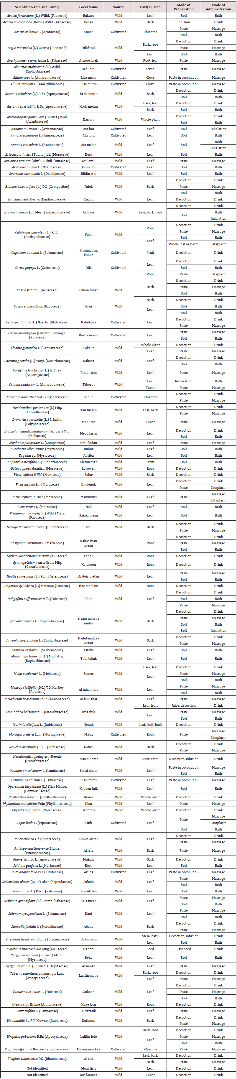
Native people in West Timor Indonesia have been exposed to malaria since long time ago. Because of this experience, it is believed that this community has developed their local concept about malaria, and how to manage it. This research was intended to document and analyze local knowledge and practices of malaria prevention and treatment developed by Tetun ethnic people in West Timor. The research was a field study, conducted through some interviews, discussions and observations. The results of this study showed that this community has long been developing various methods to prevent and threat malaria. The prevention and treatment of malaria in traditional medicine of Tetun ethnic people consists of both herbal and non-herbal methods and supported by some prohibitions and restrictions. The results also showed that the practice of traditional medicine for prevention and treatment of malaria by Tetun ethnic people can be explained scientifically. Medicinal plants that widely used like Strychnos ligustrina, Carica papaya, Momordica sp., Cleome rutidosperma, Physalis angulata, Alstonia spectabilis, Alstonia scholaris and Melia azedarach have been proven to have antimalarial activities as anti-plasmodial, antipyretic, analgesic, anti-inflammatory and immunostimulant.

Keywords: Local Knowledge; Traditional Medicine; Malaria Prevention and Treatment; Tetun Ethnic; West Timor


Introduction

Traditional communities in ancient times developed their local knowledge about the prevention and treatment of a disease based on their experience interacting with the disease for a long time. This local knowledge was then become a guidance for them to establish strategies to prevent and treat the disease, which were practiced widely in the community, and become their traditional medicine [1,2]. Traditional medicine is a term imposed on pre-scientific medical systems, and defines as a sum total of knowledge, skills and practices based on theories, beliefs and experiences of different cultural customs used in health care, disease prevention and increased physical and mental performance, which have been used for generations from one generation to the next [3,4]. Malaria is an ancient disease that has not been fully eradicated until this time [5]. Since long time ago, malaria was the main infectious disease that often attacks Timorese people, especially in Belu and Malaka Districts in West Timor (Indonesia). Several old manuscripts noted that Timorese people in early of 19th century were suffered from malaria which caused many deaths [6,7]. Until this time, Belu and Malaka Districts are still hyper-endemic areas of malaria. According to the Global Fund report, in 2014, Belu and Malaka Districts were classified as high malaria endemic areas, with the Annual Parasite Insidence (API) of 12.87o/oo and 11.58o/oo respectively, higher than Indonesian average API 1.38o/oo. Various programs for malaria prevention and eradication sponsored by the Indonesian Ministry of Health and World Health Organization such as insecticide-impregnated net, fogging, mass blood survey for early diagnosis and prompt treatment, and treat malaria patient using Artemisinin Combination Therapy (ACT) have been implemented, but decreasing of the API value is still not too convincing [8]. Cultural factors that influence public attitudes and acceptance on the programs of prevention and treatment of malaria are estimated to be one of the obstacles to the success of these programs. The implementation of various disease control programs and strategies often faces major challenges stemming from the social and cultural situation of the community. The social and cultural situation of a community in a particular place can negatively influence the choice, acceptance and use of interventions in disease control. Many programs of disease control and eradication are unsuccessful because of these social and cultural barriers. Therefore, it is very necessary to understand the local knowledge of the community, including an understanding of the health-illness concept that they believe in. An understanding of this can help policy makers in designing a sustainable and more effective disease control programs [9]. The Tetun ethnic is one of native communities that inhabit territories from the central part of Timor island (in Belu and Malaka districts, Indonesia) to the east (in Republic Democratic de Timor Lester, RDTL). Tetun people are still using traditional medicines to date, and often running various traditional medication rituals [10]. Because of their long-time interaction with malaria, it should be assumed that they have developed their own local knowledge about malaria and methods to prevent and treat it. Therefore, this research was intended to study the local knowledge of the Tetun ethnic people regarding malaria and the methods they have developed for the prevention and treatment of this disease.

Study Design

This study is a kind of research in the field of medical anthropology. This study was conducted as a qualitative exploratory research, with a field study as main technique, supported by a literature study.

Profile of Study Site and People

This research was conducted in Belu and Malaka Districts located in the central part of Timor island. These areas are located at 9°15’ S-9°34’ S and 124°40’ E-124°54’ E. Belu and Malaka are two of Indonesian territories that border directly with the Republic Democratic Timor Leste (RDTL). The topography of Belu Districts is mainly hilly, while Malaka is generally a stretch of flat land. Some areas of Malaka at the south part meet the rainy season twice in a year, while the areas of north part and also Belu areas are only have one rainy season. The main rainy season takes place between November-March due to wind that brings rain from the Indonesian Ocean. This rain occurs evenly in Malaka and Belu regions. The additional rainy season in April-June, which is limited in some areas of Malaka, is affected by wind from Australia that carries moisture from the Timor Sea. Based on the ethnolinguistics, there are four indigenous ethnic groups that live in Belu and Malaka Districts, namely Tetun, Dawan, Kemak, and Bunaq (Marae). Tetun ethnic is the majority ethnic group in Belu and Malaka, consists of approximately 80% of the population. They scaterred in almost all sub-districts of Belu and Malaka [11].

The Informants

The informants of this study were people of Tetun ethnic who have lived for long time in Belu or Malaka Districts. They were people with good knowledge and experiences of traditional medicine practices. The informants were selected through the purpossive and snowball tehniques. A total of 94 informants (42 men and 52 women) with the age of 40-90 years old were involved in this study. They came from 15 vilages of five sub-districts in Malaka (Wewiku, Malaka Barat, Weliman, Malaka Tengah and Kobalima Timur Subdistricts), and 14 vilages of ten sub-districts in Belu (Raimanuk, Tasifeto Barat, Nanaet Duabesi, Tasifeto Timur, Lasiolat, Raihat, Lamaknen, Kakuluk Mesak, Atambua Barat and Atambua Selatan Sub-districts). These informants consist of traditional public healers, home healers, and traditional medicine users.

Data Collection

Data were collected through several interviews, discussions, and observation. Interviews were conducted with a semi-structured questionnaire. Interviews were intended to collect informations about local knowledge on health-illness concept, symptoms, signs and causes of malaria, traditional methods for the prevention and treatment of malaria, and medicinal plants used for the prevention and treatment of malaria. More deep questions were developed spontaneously based on the answers given by the informants to the previous questions. Interviews and discussions were conducted in Tetun (local language) and Indonesian. We recorded the contents of every interview by wrote a detailed essence of the conversation, but not fully word by word. Several interviews were recorded with audio and video recorder. In this field study, we were assisted by several local guides to search for informants, accompanied in the interviews, to interpreted specific local terms that strange for us, and help us to search, document and collect plant specimens. All plants mentioned by informants were collected in-situ and documented by making photographs and herbaria for taxonomic identification. This field study was conducted from April 2017 to December 2017.

Data Analysis

Data obtained from interviews, discussions and observations were analyzed qualitatively, and presented in narrative or qualitative descriptions [12]. The steps of qualitative analysis are as follows:
a) Transcription of data: first of all, the interview data, discussions and field observation records were well-transcribed in a neat text.
b) Data reduction: transcripts were analyzed to marked meaningful parts, and then grouped based on the same characteristics into certain categories, i.e. the local knowledge about health-illness, local concepts about malaria, methods for the prevention and treatment of malaria, and plants used for the prevention and treatment of malaria.
c) Presentation of data: data that has been grouped were arranged regularly according to each category to make them easy to understand. Data of plants used in malaria prevention and treatment were presented in a table.
d) Verification and conclusion: determined the meaning of the data presented.

Local Concepts about Health-Illness

The concept of health and illness in Tetun community is very simple. Tetun people define health as a condition of normal, good and not sick. Illness is interpreted as a condition in which someone feels unwell or sick or has a disease in the body. Tetun traditional people state a condition as health or ill by seeing physical signs. A person is said to be health if he/she looks physically strong, fresh, agile, has a bright face and good appetite; and vice versa, if the physical performance seems weak, lethargic, pale face, lack of appetite, then the person is said to be sick or has an illness in the body. Someone is said to have recovered from illness when showing physical signs such as being able to get up, not feel dizzy anymore, being able to walk quickly and to work again, and his/her appetite is back and improved. The concept of Tetun people about health and illness is also associated with the ability to carry out daily life activities. Someone who is still able to work or move without feeling bad or pain in his body, then that person is not said to be sick. People who are clinically suffering from a certain disease but not feel sick and still able to carry out daily activities without being disturbed by the disease, then that person is not considered sick. WHO and Indonesian Ministry of Health define health as a state of complete physical, mental and social well-being, and not merely the absence of diseases or infirmity [13]. Comparing the concept of health according to Tetun people’s understanding with this official definition, it can be concluded that the concept of health of Tetun ethnic people is inadequate to describes whole condition called health, because for this community, health and illness are more related to physical performance than psychological and social performance.

Local Concept about Symptoms and Signs, and Causes of Malaria

The indigenous people of Tetun know malaria as is in mana’s (hot body, fever) with primary signs and symptoms are high fever, shivering, intermittent fever, headache, muscle and joint pain, pale, yellow eyes, and abdominal pain and/or diarrhea. Many informants did not know that swollen spleen (splenomegaly) is also one of the signs of malaria that is already severe, but they assumed that the swollen spleen can cause fever (they say “malaria”). In general, almost all the informants assumed that malaria is a common, mild and not serious disease, only a sick of hot body or fever. This local concept seems to greatly influences people’s perceptions of the danger of malaria and result in reduction of their alertness on malaria and the seriousness of managing this disease. In the local knowledge of Tetun ethnic people, the causes of malaria are: sweet food and drink, chilled, sunburn, fatigue, presence of other disease in the body, magic, cold food and drink, lack of sleep, inadequate post-natal care, spicy food, alcohol, and oily or fatty food. Tetun ethnic people assumed that sweet food and drink, sunburn, magic, spicy food, alcohol, and oily or fatty food cause an excessive heat in the body, and as a result, someone will get high fever malaria. Chilled, cold food and drink, lack of sleep is assumed to cause cold entering the body, and as the result, someone will get shivering malaria. The fatigue, presence of other disease in the body and inadequate post-natal care for mother and infant are assumed to destroy the equilibrium of hot and cold in the body and result in malaria with high fever and/or shivering. According to some informants, mosquito as malaria transmitter was a new knowledge that coming from outside, introduced by the Catholic missionaries from Europe. According to Foster dichotomous on causes of disease [14], the causes of malaria in the local concept of Tetun people are naturalistic, not personalistic. Factors such as sweet foods or drinks, long time in rain, water or cold places, long working under the hot sun, fatigue and the presence of other diseases in the body are naturalistic properties that cause heat-cold balance in the human body to be disrupted, and then causes someone to get malaria. Many Tetun people do not consider mosquito as carrier of malaria, causing them to have low awareness of the threat of mosquitoes. This may be one of the causes of the still high endemic of malaria in Belu and Malaka until this time [15].

Methods for The Prevention and Treatment of Malaria

The Tetun ethnic people have their own patterns or habits of life that they do for generations to prevent malaria attacks. The methods that are considered effective in preventing attacks of malaria are: luli or hale’u, drink medicinal concoction of bitter herbs, eat bitter food, and drink tua moruk. Luli or hale’u means avoiding things that can cause malaria (according to their local concepts about the cause of malaria), which are: not eating sweets frequently, not working for long time under the rain or hot sun, and not too tired at work or physical activities. Eating bitter foods, especially papaya and bitter melon, and drinking bitter palm sap tua moruk are also considered effective to prevent someone from being attacked by malaria. Some informants who previously linked malaria with mosquitoes stated that repelling mosquitoes using smoke of burned aromatic plants and sleeping under mosquito nets are effective for malaria prevention. The treatment of malaria in traditional medicine of Tetun ethnic consists of herbal and non-herbal methods. Herbal method consists of drinking herbal concoction, inhaling the vapor of boiled medicinal plant, massage with paste of medicinal plant, bath with water of boiled medicinal plant, and attach the paste of medicinal plant as a cataplasm on the swollen spleen. A non-herbal method is sunu kok, that is burning the waist above the swollen spleen using a piece of coconut shell coal or a heated metal. The results of the interviews showed that most traditional medication for malarial patient usually combine two or more methods. It was found also that the role of traditional healer in the treatment of malaria patient is not so important. Tetun ethnic people assumed that malaria is a common and not a serious disease, thus the treatment of malaria does not require a high competency healer. Several informants stated that they usually conducted self- and home-medication for malaria complaint. In the traditional medicine of Tetun ethnic people, the treatment of malaria is a simple treatment for reducing heat or fever [15]. The assumption of malaria as a common, mild and not a serious disease results in lack of awareness about dangers of malaria. It was found that in many cases, health workers often complain of disobedience of patients who stop taking antimalarial drugs immediately after they feel cured (being able to get up, not feel dizzy anymore, being able to work again, and the appetite is improved), even though Plasmodium in their blood has not been completely eliminated. As the result, the success of the malaria eradication program in this area has increased very slowly [8].

Plants Used for The Prevention of Malaria

Tetun ethnic people believe that consumption of bitter food or drink can prevent someone from malaria attacks. Therefore, small children are often forced by their parents to eat stew and drink decoctions of flowers, leaves and young fruit of Carica papaya, or young fruit of Momordica sp. (M. charantia or M. balsamina). Some informants gave information that if they feel tired, achy and lack of appetite, they will drink decoction of Carica papaya leaves, fruit of Momordica charantia, Melia azadarach leaves, Alstonia scholaris, Alstonia spectabilis or Strychnos ligustrina stem bark. Consumption of these plants’ decoction is believed to restoring body freshness, increasing appetite, eliminating fatigue, and thus, preventing from malaria attack. Some informants also believed that drinking tua moruk is effective in malaria preventon. Tua moruk is a traditional drink made by fresh tapped palm sap soaked with the stem bark of Alstonia scholaris, Alstonia spectabilis or Strychnos ligustrina soaked in it. Several publications of other previous studies showed that the bitter plants used by Tetun people to prevent malaria has been shown to have pharmacological activities as antiplasmodium and immunostimulant [16-18].

Plants Used for The Treatment of Malaria

In this study, we recorded a total of 96 species from 39 families used by Tetun people in various formula for drink, massage, bath, inhalation and cataplasm (Table 1). Strychnos ligustrina, Carica papaya, Cleome rutidosperma, Physalis angulata, Alstonia spectabilis, Alstonia scholaris and Melia azedarach are some of the most widely plants used in various formula for drink. For massage, Garuga floribunda, Jatropha curcas, Acorus calamus, Allium cepa, Drynaria quercifolia, Ocimum sp. and Ruta graveolens are common. For bathing, people use Tamarindus indica, Psidium guajava, Melicope latifolia and Blumea balsamifera. Leaves of Brucea javanica, Annona muricata and Annona reticulata are used in inhalation method. Root of Moringa oleifera and leaves of Ficus hispida are used as cataplasm to reduce the swollen spleen [19]. Several plants were found in various formula for more than one mode of application. Several previous publications showed that most of these plants are also used in other traditional medicine for the same purpose in many areas of Indonesia and the world [16,18], and have been scientifically proven to have pharmacological activities as true antimalarial (antiplasmodial) and/or indirect antimalarial such as antipyretic, analgesic, anti-inflammatory and immunostimulant [20].
Table 1: Plants used by Tetum ethnic people for the treatment of malaria.

Lupinepublishers-openaccess-complementary-alternative-medicine-journal


Conclusion

The practice of preventing and treating malaria in the traditional medicine of Tetun ethnic people is a direct implementation of their local knowledge about malaria. The local concept of signs and symptoms and the causes of malaria encourage traditional people to create methods to prevent and treat malaria. The local concept of the Tetun ethnic people about malaria is the main reference in the creation of rules regarding prohibitions and restrictions, and recommendations for preventing attacks of malaria. The local concept of the causes of malaria determines the choice of plants for the treatment of malaria. Scientifically, these plants have been proven to have activities as true antimalarial and indirect antimalarial. The local concept of malaria as a common, mild and harmless disease causes that the role of traditional healer is not always needed in the treatment of malaria. Methods for the prevention and treatment of malaria developed by Tetun ethnic people consist of both herbal and non-herbal methods and supported by the implementation of several prohibitions and restrictions to provide healing for the sufferers of malaria.


Acknowledgement

We thank to Indonesian Ministry of Research, Technology and Higher Education, for financial support (Research Contract No. 0668/K8/KM/2018).

For More Lupine Publishers Open Access Journals Please visit our Website:

To Know More About Open Access Publishers Please Click on Lupine Publishers

Follow on Twitter:  https://twitter.com/lupine_online

Thursday, March 12, 2020

Smoking May Not Increase Systolic and Diastolic Blood Pressures

  Abstract Background: We tried to understand whether there are some effects of smoking on systolic and diastolic blood pressure...